. 2025 Jun 20;53(12):gkaf543.
doi: 10.1093/nar/gkaf543.

Pyridine-bis(benzimidazole) induces DNA damage at G-quadruplex loci and promotes synthetic lethality with DNA repair inhibition

Affiliations

Pyridine-bis(benzimidazole) induces DNA damage at G-quadruplex loci and promotes synthetic lethality with DNA repair inhibition

Jiangyu Yan et al. Nucleic Acids Res. .

Abstract

G-quadruplexes (G4s) are noncanonical DNA structures that play key roles in regulating replication, transcription, and genome stability. Here, we investigate the effects of pyridine-bis(benzimidazole) (PyBI), a selective parallel and hybrid G4 stabilizer, on genome stability in cells. Biophysical and biochemical assays confirm PyBI's strong affinity for parallel G4s, leading to replication fork stalling and transcriptional repression of G4-associated oncogenes. Cleavage under targets and tagmentation (CUT&Tag) sequencing reveals a PyBI-induced genome-wide increase in G4 peaks, particularly at promoter and transcription start site regions. This G4 induction and stabilization triggers replication stress, G2/M arrest, and apoptosis. DNA repair pathway profiling shows that PyBI-induced G4 stabilization activates both homologous recombination and nonhomologous end joining (NHEJ), with a predominant role for NHEJ, as indicated by higher 53BP1 colocalization at G4 sites. Genome-wide mapping of PyBI-induced DNA breaks further supports a direct link between G4 stabilization and DNA damage. Moreover, PyBI shows synthetic lethality in DNA repair-deficient contexts, highlighting its therapeutic potential. These findings provide mechanistic insights into the genotoxic effects of PyBI and highlight its potential as a novel anticancer agent targeting G4-mediated genome instability.


Conflict of interest statement

None declared.

Figures

Graphical Abstract
Graphical Abstract
Figure 1.
Figure 1.
PyBI is a G4 stabilizer. (A) Chemical structure of the PyBI. (B) Dissociation constants (Kd) illustrating PyBI’s interaction with G4 and non-G4 sequences. (C) In vitro FRET melting assay comparing PyBI’s effect on G4-forming DNA oligonucleotides versus a non-G4 forming hairpin control. (D) Schematic depiction of PyBI hindering DNA polymerase progression by stabilizing G4 structures. (E) Half-inhibitory concentration (IC50) of PyBI causing DNA polymerase arrest, PT (prematurely terminated), and FL (full-length product). The presented data represent the mean values of three independent experiments, with error bars indicating the SD.
Figure 2.
Figure 2.
PyBI increases intracellular DNA G4. (A) BG4 foci images of cells treated with different concentrations (1, 2, and 5 μM) of PyBI for 2, 6, and 24 h. (B) Quantitative analysis of BG4 foci per nucleus in panel (A). (C) Representative images of HeLa cells treated with PyBI (1 μM) and PDS (10 μM) for 24 h. (D) Quantitative analysis of BG4 foci per nucleus. The error bars represent the mean ± SD from three independent experiments. At least 300 cells were counted. Statistical significance was determined using a paired t-test. *P < .05, **P < .01, ***P < .001, and ****P < .0001; ns: not significant (P > .05). Scale bars, 10 μm.
Figure 3.
Figure 3.
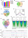
PyBI increases intracellular DNA G4. (A) Percentage of G4P and BG4 peaks overlapping with PQS. (B) The overlap of peaks between Ctrl G4P and PyBI G4P. (C) Distribution of enhanced G4P peaks in genomic regions. (D) Profiling of increased G4P sites concentrated in the TSS ± 1 Kb region after PyBI treatment. (E) The overlap of peaks between Ctrl BG4, PyBI BG4, and PDS BG4 peaks. (F) Profile (top) and heatmap (bottom) of Ctrl and PyBI BG4 at the TSS ± 1 kb regions. (G) Genomic tracks comparing PyBI-enhanced G4 and PDS-enhanced G4 versus control G4 (all genomic tracks display the average of two duplicate samples).
Figure 4.
Figure 4.
PyBI stabilizes G4, leading to replication fork stalling and DNA damage. (A) Upper: Schematic illustration of the fiber patterns. Cells were pulse-labeled with CldU (green) for 20 min, followed by a 20-min pulse of IdU (red) combined with 5 μM PyBI. Lower: Representative DNA fiber immunofluorescence images from cells treated with or without PyBI. (B) Quantitation of DNA fibers displaying ongoing forks, replication stalling, or new origins. At least 150 cells were counted for DNA fiber sample. (C) γ-H2AX and BG4 foci images of cells treated with 5 μM PyBI. (D) Quantitative analysis of γ-H2AX foci per nucleus. At least 300 cells were counted. (E) The percentage of γ-H2AX foci colocalized with BG4 foci in HeLa cells treated with 5 μM PyBI. (F) Alkaline comet assay monitoring DNA damage. Representative images of HeLa cells treated with PyBI (5 μM) for 24 h. (G) Statistical analysis of DNA comet tail and tail moment. Data in all panels are from at least two biological replicates, and in each experiment an average of 180 cells per sample was determined. Statistical significance was determined using a paired t-test. *P < .05, **P < .01, ***P < .001, and ****P < .0001; ns: not significant (P > .05). Scale bars, 10 μm.
Figure 5.
Figure 5.
PyBI induces DNA damage and depends on HR and NHEJ repair pathways. (A) RAD51 and BG4 foci images of cells treated with 5 μM PyBI. (B) Quantitative analysis of RAD51 foci per nucleus in panel (A). (C) The percentage of RAD51 foci colocalized with BG4 foci in HeLa cells treated with 5 μM PyBI. (D) 53BP1 and BG4 foci images of cells treated with 5 μM PyBI. (E) Quantitative analysis of 53BP1 foci per nucleus in panel (D). (F) The percentage of 53BP1 foci colocalized with BG4 foci in HeLa cells treated with 5 μM PyBI. Data are presented as mean ± SD from three independent experiments. At least 300 cells were counted. Statistical significance was determined using a paired t-test. *P < .05, **P < .01, ***P < .001, and ****P < .0001; ns: not significant (P > .05). Scale bars, 10 μm.
Figure 6.
Figure 6.
PyBI (5 μM)-induced DNA damage is mediated by G4. (A) Illustration of CUT&Tag assay with RAD51 and 53BP1. (BC) RAD51 and 53BP1 profiles at G4P peak ± 1 kb regions. (D) Venn diagram showing overlap between RAD51 and G4 peaks. (E) Overlap between 53BP1 and G4 peaks. (F) The overlap between PyBI-induced DSB and G4 peaks. (G) Genomic tracks depicting increased RAD51 and 53BP1 sites after PyBI treatment, coinciding with enhanced G4 sites (Average of two duplicate samples).
Figure 7.
Figure 7.
Synthetic lethality of PyBI in the context of HR and NHEJ pathway deficiencies. (A) Cell viability of HeLa cells transfected with siRAD51 or siKU80 following treatment with PyBI for 24 h. (B) Synergy analysis of PyBI in combination with RI-1 (RAD51 inhibitor) and PIK-90 (DNA-PK inhibitor) in HeLa cells after 24 h of treatment. (CD) Immunofluorescence staining and quantification of γ-H2AX foci in HeLa cells transfected with siRAD51 or siKU80. (EF) Immunofluorescence staining and quantification of γ-H2AX foci in HeLa cells treated with PyBI in combination with RI-1 or PIK-90. Data are presented as mean ± SD from three independent experiments. Statistical significance was determined using a paired t-test. *P < .05, **P < .01, ***P < .001, and ****P < .0001. Scale bars, 10 μm.

Similar articles

References

    1. Moye AL, Porter KC, Cohen SB et al. Telomeric G-quadruplexes are a substrate and site of localization for human telomerase. Nat Commun. 2015; 6:7643. 10.1038/ncomms8643. - DOI - PMC - PubMed
    1. Wang KB, Elsayed MSA, Wu G et al. Indenoisoquinoline topoisomerase inhibitors strongly bind and stabilize the MYCpromoter G-quadruplex and downregulate MYC. J Am Chem Soc. 2019; 141:11059–70. 10.1021/jacs.9b02679. - DOI - PMC - PubMed
    1. Besnard E, Babled A, Lapasset L et al. Unraveling cell type-specific and reprogrammable human replication origin signatures associated with G-quadruplex consensus motifs. Nat Struct Mol Biol. 2012; 19:837–44. 10.1038/nsmb.2339. - DOI - PubMed
    1. Varshney D, Spiegel J, Zyner K et al. The regulation and functions of DNA and RNA G-quadruplexes. Nat Rev Mol Cell Biol. 2020; 21:459–74. 10.1038/s41580-020-0236-x. - DOI - PMC - PubMed
    1. Mendoza O, Bourdoncle A, Boule JB et al. G-quadruplexes and helicases. Nucleic Acids Res. 2016; 44:1989–2006. 10.1093/nar/gkw079. - DOI - PMC - PubMed

MeSH terms

LinkOut - more resources