. 2025 Jun 20;53(12):gkaf562.
doi: 10.1093/nar/gkaf562.

Structural and functional studies of the main replication protein NS1 of human parvovirus B19

Affiliations

Structural and functional studies of the main replication protein NS1 of human parvovirus B19

Yixi Zhang et al. Nucleic Acids Res. .

Abstract

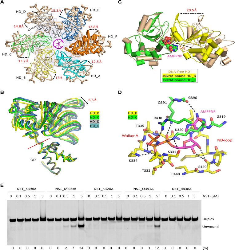
Parvovirus B19 (B19V) is a ubiquitous virus that can infect the majority of human population and cause erythema infectiosum, acute arthropathy, and many other diseases. The main replication protein NS1 plays a critical role in cell cycle arrest, transactivation of viral and host genes, and replication and package of B19V genome. Both DNA nicking and unwinding activities are required for the in vivo function of NS1, but the underlying basis is poorly understood. Here, we report extensive structural and biochemical studies of NS1, showing that NS1 can unwind various types of DNA substrates. The cryo-electron microscopy (cryo-EM) structures reveal the detailed mechanisms for ATP binding and hydrolysis, and DNA binding and unwinding by NS1. In addition to the SF3 HD domain, the C-terminal region is also required for double-stranded DNA (dsDNA) nicking by NS1. Unexpectedly, instead of enhancing, the dsDNA nicking activity of NS1 is negatively regulated by its DNA unwinding ability, suggesting that they likely function in different stages. This study advances our understanding of the structure and function of NS1 and other parvoviral replication proteins, such as the Rep proteins of adeno-associated virus.


Conflict of interest statement

None declared.

Figures

Graphical Abstract
Graphical Abstract
Figure 1.
Figure 1.
Domain architecture and DNA unwinding activity of B19V NS1 protein. (A) Domain architecture of B19V NS1 and the homologous proteins of AAV. (B) In vitro DNA unwinding assays catalyzed by NS1_2-570 protein. The detailed sequences of the four DNA substrates are available in Supplementary Fig. S3. The substrate unwinding percentage (%) is shown at the bottom of the gels. Experiments were repeated independently three times with similar results. (C) Overall folding and assembly of NS1 in the NS1_2-570/AMPPNP structure. A schematic view showing the assembly of NS1 dodecamer is shown at the bottom. In the left panel, the 12 NS1_2-570 protomers are shown as cartoon in different colors. In the right panel, the protomers in the upper hexamer are shown as cartoon, whereas the protomers in the lower hexamer are shown as surface in white. AMPPNP molecules are shown as red spheres.
Figure 2.
Figure 2.
AMPPNP recognition in the NS1_2-570/AMPPNP structure. (A) Density maps of AMPPNP and the surrounding residues. (B) The detailed interactions between AMPPNP and NS1_2-570. (C) Stick-and-surface presentation showing the open conformation of the AMPPNP-binding pocket. (D) Superposition of AMPPNP and ADP bound in the NS1_2-570/AMPPNP structure and the AAV2 Rep40/ADP complex (PDB_ID: 1U0J), respectively. (E) In vitro assays showing the effects of mutation of AMPPNP-interacting residues on duplex DNA-1 unwinding by NS1. The substrate unwinding percentage (%) is shown at the bottom of the gels. Experiments were repeated independently three times with similar results. For the Rep40/ADP complex, ADP and Rep40 are colored in cyan and gray, respectively. For the NS1_2-570/AMPPNP structure, AMPPNP is shown as sticks in atomic color (C, yellow; N, blue; O, red; P, orange). Mg2+ and water molecules are shown as spheres in black and red, respectively.
Figure 3.
Figure 3.
ssDNA binding by NS1_2-570. (A) The final density maps of the NS1_2-570/ssDNA/AMPPNP structure. (B) The overall conformation and assembly of the NS1_2-570/ssDNA/AMPPNP structure. (C) The detailed interactions between ssDNA and NS1_2-570. The C-atoms of the ssDNA are colored magenta. The C-atoms of the NS1_2-570 protomers A–F are colored in cyan, yellow, green, gray, light blue, and orange, respectively.
Figure 4.
Figure 4.
Conformational changes induced by ssDNA binding. (A) Superposition of DNA-free and ssDNA-bound NS1_2-570 structures, which are presented in sausage views based on the B factors. (B) Superposition of NS1_2-570 protomers B–E in the ssDNA-bound structure. (C) Superposition showing the shifting of the neighboring SF3 HD domain. (D) The detailed interactions between AMPPNP and NS1_2-570 in the ssDNA-bound structure. (E) In vitro assays showing the effects of the ssDNA- or AMPPNP-interacting residue mutations on duplex DNA-1 unwinding by NS1. The substrate unwinding percentage (%) is shown at the bottom of the gels. Experiments were repeated independently three times with similar results. The NS1_2-570 protomers are colored in pink in the DNA-free structure. For the ssDNA-bound structure, the C-atoms of protomers A–F are colored in cyan, yellow, green, gray, light blue, and orange, respectively. The C-atoms of AMPPNP are colored in magenta.
Figure 5.
Figure 5.
dsDNA binding by NS1_2-570. (A) The final density maps of the NS1_2-570/dsDNA/AMPPNP structure. (B) The overall conformation and assembly of the NS1_2-570/dsDNA/AMPPNP structure. (CD) The detailed interactions between dsDNA and NS1_2-570 observed in the NS1_2-570/dsDNA/AMPPNP structure. (E) In vitro assays showing the effects of mutation of dsDNA-interacting residues on duplex DNA-1 unwinding by NS1. The substrate unwinding percentage (%) is shown at the bottom of the gels. Experiments were repeated independently three times with similar results. The C-atoms of dsDNA are colored in magenta. The C-atoms of protomers A–F are colored in cyan, yellow, green, gray, light blue, and orange, respectively.
Figure 6.
Figure 6.
In vitro dsDNA cleavage assays. (A) Duplex DNA-1 cleavage by WT NS1_2-570 and mutants with the dsDNA-interacting residues mutated. (B) Duplex DNA-1 cleavage by WT NS1_2-570 and mutants with the ssDNA-interacting residues mutated. (CD) Duplex DNA-1 cleavage by NS1_2-570 mutants with the ATP-interacting residues mutated. The substrate cleavage percentage (%) is shown at the bottom of the gels. Experiments were repeated independently three times with similar results.
Figure 7.
Figure 7.
Correlation between DNA unwinding and DNA cleavage activities of NS1. (A) Comparison of in vitro dsDNA cleavage activity of NS1_2-570 in the presence or absence of ATP. (B) Comparison of in vitro duplex DNA-1 cleavage activity of NS1_2-570 in the presence or absence of AMPPNP. (C) Sequences of duplex DNA-1, duplex DNA-2, and duplex DNA-3. (D) In vitro dsDNA unwinding by NS1_2-570. The substrate cleavage percentage (%) is shown at the bottom of the gels. Experiments were repeated independently three times with similar results.
Figure 8.
Figure 8.
Identification of regions important for the DNA cleavage activities of NS1. (A) The close-up view showing the density maps for the potential NS1_Nuc domain observed in the NS1_2-570/dsDNA/AMPPNP structure. (B) Comparison of in vitro duplex DNA-1 cleavage activity of NS1_2-570, NS1_2-176, and NS1_200-570. (C) Sequences of the 501–570 region in WT NS1_2-570 and the DE501-522A mutant protein. (D) Comparison of duplex DNA-1 cleavage activity of NS1_2-501, NS1_2-522, and NS1_2-530 mutants. (E) Comparison of duplex DNA-1 cleavage activity of WT and the DE501-522A mutant proteins of NS1_2-570. All assays were done in the absence of ATP. The substrate cleavage percentage (%) is shown at the bottom of the gels. Experiments were repeated independently three times with similar results.

Similar articles

References

    1. Cossart YE, Cant B, Field AM et al. Parvovirus-like particles in human sera. Lancet. 1975; 1:72–3. 10.1016/S0140-6736(75)91074-0. - DOI - PubMed
    1. Young NS, Brown KE Mechanisms of disease—parvovirus B19. N Engl J Med. 2004; 350:586–97. 10.1056/NEJMra030840. - DOI - PubMed
    1. Wurdinger M, Modrow S, Plentz A Impact of parvovirus B19 viremia in liver transplanted children on anemia: a retrospective study. Viruses. 2017; 9:149. 10.3390/v9060149. - DOI - PMC - PubMed
    1. Pinto NC, Newman C, Gomez CA et al. Parvovirus B19-induced severe anemia in heart transplant recipients: case report and review of the literature. Clin Transplant. 2019; 33:e13498. 10.1111/ctr.13498. - DOI - PMC - PubMed
    1. Seng C, Watkins P, Morse D et al. Parvovirus B19 outbreak on an adult ward. Epidemiol Infect. 1994; 113:345–53. 10.1017/S0950268800051773. - DOI - PMC - PubMed

MeSH terms

LinkOut - more resources