. 2025 Jul 22;10(7):e0060325.
doi: 10.1128/msystems.00603-25. Epub 2025 Jun 26.

Extremely distinct microbial communities in closely related leafhopper subfamilies: Typhlocybinae and Eurymelinae (Cicadellidae, Hemiptera)

Affiliations

Extremely distinct microbial communities in closely related leafhopper subfamilies: Typhlocybinae and Eurymelinae (Cicadellidae, Hemiptera)

Michał Kobiałka et al. mSystems. .

Abstract

Among the Hemiptera insects, a widespread way of feeding is sucking sap from host plants. Due to their nutrient-poor diet, these insects enter into obligate symbiosis with their microorganisms involved in the synthesis of components essential for host survival. However, within the Cicadellidae family, there is a relatively large group of mesophyll feeders-Typhlocybinae-that is considered to be devoid of obligate symbiotic companions. In this work, we examine the composition of microorganisms in this subfamily and compare the results with their close relatives-the Eurymelinae subfamily. To study the microbiome, we used high-throughput next-generation sequencing (NGS, Illumina) and advanced microscopic techniques, such as transmission electron microscopy (TEM) and fluorescence in situ hybridization (FISH), in a confocal microscope. In the bodies of Typhlocybinae insects, we did not detect the presence of microorganisms deemed to be obligate symbionts. Their microbial communities consist of facultative symbionts, mainly alphaproteobacteria such as Wolbachia or Rickettsia as well as others that can be considered as facultative, including Spiroplasma, Acidocella, Arsenophonus, Sodalis, Lariskella, Serratia, Cardinium, and Asaia. On the other hand, the Eurymelinae group is characterized by a high diversity of microbial communities, both obligate and facultative, similar to other Cicadomorpha. We find co-symbionts involved in the synthesis of essential amino acids such as Karelsulcia, betaproteobacteria Nasuia, or gammaproteobacteria Sodalis. In other representatives, we observed symbiotic yeast-like fungi from the family Ophiocordycipitaceae or Arsenophonus bacteria inhabiting the interior of Karelsulcia bacteria. Additionally, we investigated some aspects of symbiont transmission and the phylogeny of symbiotic organisms and their hosts.

Importance: The Typhlocybinae and Eurymelinae leafhoppers differ significantly in their symbiotic communities. They have different diets, as Typhlocybinae insects feed on parenchyma, which is richer in nutrients, while Eurymelinae, like most representatives of Auchenorrhyncha, consume sap from the phloem fibers of plants. Our work presents comprehensive studies of 42 species belonging to the two above-mentioned, and so far poorly known, Cicadomorpha subfamilies. Phylogenetic studies indicate that the insects from the studied groups have a common ancestor. The diet shift in the Typhlocybinae leafhoppers contributed to major changes in the composition of microorganisms inhabiting the body of these insects. Research on the impact of diet on the microbiome and the subsequent consequences on the evolution and adaptation of organisms plays an important role in the era of climate change.

Keywords: endosymbionts; insect microbiome; leafhoppers; microbial ecology; symbiotic bacteria; symbiotic fungi.


Conflict of interest statement

The authors declare no conflict of interest.

Figures

Fig 1
Fig 1
Eurymelinae symbiotic systems, 16S rRNA metabarcoding. (A) Cladogram based on Bayesian analysis of the most abundant sequences of the bacteria Karelsulcia for each individual, showing phylogeny among Eurymelinae species. Posterior probabilities are shown above the nodes. The outgroup is Karelsulcia from Centrotus cornutus (MN082139.1) (50). Below the cladogram, the graphs show the percentage of the relative abundance of bacteria for each sample based on the number of sequence reads. (B) Relative abundance of OTUs of both bacterial and fungal microorganisms, collectively for each Eurymelinae species.
Fig 2
Fig 2
Eurymelinae symbiotic systems. (A) A. ribauti, bacteriome containing Karelsulcia bacteria (green), surrounded by a fat body where yeast-like symbionts of the genus Hirsutella are present (red). (B) M. vicina, bacteriome containing Karelsulcia bacteria (green), surrounded by a fat body where yeast-like symbionts of the genus Ophiocordyceps (red) are present. (C) M. prasina, Ophiocordyceps yeast-like symbionts (y) in insect fat body. (D) P. tiliae, Karelsulcia bacteria (s) in bacteriocyte; note Wolbachia bacteria (white arrowheads) inside the cell nucleus (nc). (E and F) I. stigmaticalis, Arsenoponus bacteria (purple) residing inside Karelsulcia bacteria (green); Nasuia bacteria (red) present in bacteriocytes forming a separate zone. (G) B. larvatus, Arsenoponus bacteria (a) infecting Karelsulcia bacteria (s). (H) I. stigmaticalis, bacteriocytes filled with Karelsulcia bacteria (s) infected by Arsenophonus bacteria (a); in the upper left corner, a fragment of a bacteriocyte occupied by Nasuia bacteria (n). (I) P. albicans and (J) P. populi, bacteriome composed of two zones—bacteriocytes filled with Karelsulcia bacteria (green) and Nasuia bacteria (red). (K) P. albicans and (L) P. populi, pleomorphic Karelsulcia bacteria inhabiting bacteriocytes. (M) P. albicans, the fragment of a bacteriocyte filled with pleomorphic Nasuia bacteria (n). (N) P. confusus, bacteriocytes filled with Karelsulcia bacteria (green) infected by Arsenophonus bacteria (purple). (O and P) P. confusus, Karelsulcia bacteria (s) filled with many rod-shaped Arsenophonus bacteria (a); in the upper left corner, the fragment of a bacteriocyte occupied by Nasuia bacteria (n). (R) P. nitidissimus, two zones of a bacteriome: pleomorphic Karelsulcia bacteria (s) (left) and rod-shaped Sodalis bacteria (sod) (right). (S) T. tremulae and (T) V. ustulatus, bacteriome where bacteriocytes filled with various bacteria form three distinct zones: Karelsulcia (green), Sodalis (yellow), and Nasuia (red). (U) T. distinguendus, pleomorphic Karelsulcia bacteria (s) inhabiting bacteriocytes; note rod-shaped alphaproteobacteria present in the cell nucleus (white arrowheads). (W) T. distinguendus, two zones of bacteriome: rod-shaped Sodalis bacteria (sod) (left) and pleomorphic Nasuia bacteria (n) (right); note the viruses (v) residing in the bacteriome epithelium between zones. (V) V. ustulatus, two zones of bacteriome: pleomorphic Karelsulcia bacteria (s) (left) and rod-shaped Sodalis bacteria (sod) (right). (X) O. alni, pleomorphic Nasuia bacteria (n) inhabiting bacteriocytes; note rod-shaped alphaproteobacterial present in the cell nucleus (white arrowheads). (Y) O. flavicollis, bacteriocytes filled with Karelsulcia (green) and Nasuia (red) bacteria forming two separate bacteriomes. (A, B, E, F, I, J, N, S, T, Y) Confocal microscope; cell nuclei, DAPI staining (blue); scale bar: 40 µm. (C, D, G, H, K–M, O–R, U–X) TEM; be, bacteriome epithelium; black arrowheads, rod-shaped gammaproteobacterium; l, lipid droplet; lb, lamellar body; m, mitochondrion; nc, cell nucleus; white arrowhead, rod-shaped alphaproteobacterium; scale bar: 3 µm.
Fig 3
Fig 3
Facultative bacteria in various tissues of Typhlocybinae and Eurymelinae insects. (A) P. populi and (B) P. albicans, Wolbachia bacteria (red) present in Nasuia bacteriocytes zone. (C) V. ustulatus, alphaproteobacteria (white arrowheads) occurring in the cytoplasm and nucleus (nc) of Karelsulcia (s) bacteriocyte. (D) E. aurata, Wolbachia bacteria (red) present in the midgut epithelium. (E) F. citrinella, alphaproteobacteria (white arrowheads) settling the cell of the midgut epithelium. (F) E. lethierryi, gammaproteobacteria (black arrowheads) present in the midgut epithelium. (G) P. albicans and (H) V. ustulatus, Wolbachia bacteria (red) occurring in the midgut epithelium. (I) Z. hyperici (J), V. ustulatus (K), T. quercus (L), and M. vicina, alphaproteobacteria (white arrowheads) present in the midgut epithelium. (M) E. calcarata, Wolbachia bacteria (red) in insect fat body. (N) F. citrinella, alphaproteobacteria (white arrowheads) settling in fat body cells. (O) P. albicans, Wolbachia bacteria (red) occurring in trophocytes (ovary). (P) T. tremulae, alphaproteobacteria (white arrowheads) settling in the nucleus of fat body cells. (R) P. tiliae, alphaproteobacteria (white arrowheads) in trophocytes (tr) (ovary); note yeast-like microorganism (y) in the fat body, lower right corner. (A, B, D, G, H, M, O) Confocal microscope; cell nuclei, DAPI staining (blue); scale bar: 40 µm. (C, E, F, I-L, N, P, R) TEM; l, lipid droplet; m, mitochondrion; mv, intestinal microvilli; nc, cell nucleus; y, yeast-like microorganism; scale bar: 3 µm.
Fig 4
Fig 4
Typhlocybinae microorganisms composition and its comparison to Eurymelinae microbial system. (A) The percentage relative abundance of bacteria for Typhlocybinae species based on the number of sequence reads. (B) The PCA analysis of Typhlocybinae and Eurymelinae microbiome diversity; the arrows indicate the loadings—the most important types of bacteria differentiating two groups of insects. (C) The PCA analysis of Eurymelinae microbiome diversity for various plant subfamilies of which the insects feed; the arrows indicate the loadings—the most important types of bacteria differentiating host plant groups. (D) The PCA analysis of Typhlocybinae and Eurymelinae microbiome diversity for various plant subfamilies of which the insects feed; the arrows indicate the loadings—the most important types of bacteria differentiating host plant groups.
Fig 5
Fig 5
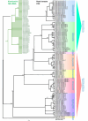
Cladograms based on Bayesian analysis of bacteria Karelsulcia 16S rDNA and COI insect gene, showing cophylogeny between Eurymelinae and their Karelsulcia symbiont (left side) and phylogeny between both Typhlocybinae and Eurymelinae species (right side). Posterior probabilities are present above the nodes. Outgroup Karelsulcia of Centrotus cornutus (MN082139.1) (50); for COI – C. cornutus (MW536003.1) (51).
Fig 6
Fig 6
Transovarial transmission of bacteria in Eurymelinae leafhoppers. (A) M. vicina, Karelsulcia bacteria (green) and Ophiocrodyceps yeast-like symbionts (red) infecting follicular cells (fc) surrounding the posterior pole of the oocyte (oc). (B) P. albicans, Karelsulcia (green) and Nasuia (red) bacteria infecting follicular cells (one of them marked by a dashed line) and forming cap-like “symbiont ball” (dotted line) in an invagination of oolemma. (C) B. larvatus, the fragment of follicular cells (fc) filled with Karelsulcia (s) and Nasuia (n) bacteria; note Arsenophonus bacteria (a) inside Karelsulcia (s). (D) P. albicans, 3D view of a “symbiont ball” consisted of Karelsulcia (green) and Nasuia (red); note the faint green and red flashes of autofluorescence on the chorion. (E) T. tremulae, the three types of bacteria: Karelsulcia (s), Sodalis (sod), and Nasuia (n) present in follicular cells. (F) M. prasina, the fragment of a “symbiont ball” consisted of Karelsulcia bacteria (s) and Ophiocordyceps yeast-like symbionts (y); note follicular cell (fc) in the bottom left corner. (G) V. ustulatus, the fragment of a “symbiont ball” consisted of Karelsulcia (s), Sodalis (sod), and Nasuia (n) bacteria. (H) B. larvatus, the fragment of a “symbiont ball” consisted of Karelsulcia (s) and Nasuia (n) bacteria; note Arsenophonus bacteria (a) inside Karelsulcia (s); note the fragment of an oocyte (oc) in the upper left corner. (A, B, D) Confocal microscope; cell nuclei, DAPI staining (blue); oc, oocyte; scale bar: 40 µm. (C, E, F through H) TEM, nc, follicular cell nucleus; scale bar: 3 µm.

Similar articles

References

    1. Cryan JR, Urban JM. 2012. Higher-level phylogeny of the insect order Hemiptera: is Auchenorrhyncha really paraphyletic? Syst Entomol 37:7–21. doi: 10.1111/j.1365-3113.2011.00611.x - DOI
    1. Dietrich CH, Allen JM, Lemmon AR, Lemmon EM, Takiya DM, Evangelista O, Walden KKO, Grady PGS, Johnson KP. 2017. Anchored hybrid enrichment-based Phylogenomics of leafhoppers and treehoppers (Hemiptera: Cicadomorpha: Membracoidea). Insect Syst Divers 1:57–72. doi: 10.1093/isd/ixx003 - DOI
    1. Zahniser JN, Dietrich CH. 2010. Phylogeny of the leafhopper subfamily Deltocephalinae (Hemiptera: Cicadellidae) based on molecular and morphological data with a revised family-group classification. Syst Entomol 35:489–511. doi: 10.1111/j.1365-3113.2010.00522.x - DOI
    1. Dietrich CH. 2013. South American leafhoppers of the tribe Typhlocybini (Hemiptera: Cicadellidae: Typhlocybinae). Zoologia (Curitiba) 30:519–568. doi: 10.1590/S1984-46702013000500008 - DOI
    1. Cao Y, Dietrich CH, Kits JH, Dmitriev DA, Richter R, Eyres J, Dettman JR, Xu Y, Huang M. 2023. Phylogenomics of microleafhoppers (Hemiptera: Cicadellidae: Typhlocybinae): morphological evolution, divergence times, and biogeography. Insect Syst Divers 7. doi: 10.1093/isd/ixad010 - DOI

LinkOut - more resources