

Characterization of the Core Temperature Response of Free-Moving Rats to 1.95 GHz Electromagnetic Fields
- PMID: 40569088
- PMCID: PMC12199690
- DOI: 10.1002/bem.70013
Characterization of the Core Temperature Response of Free-Moving Rats to 1.95 GHz Electromagnetic Fields
Abstract
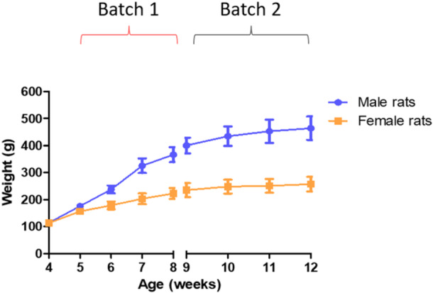
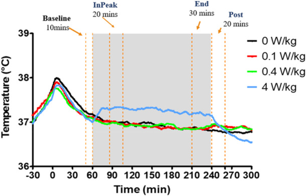
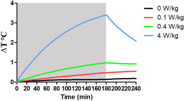
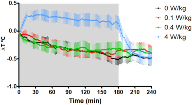
The present study investigated the core body temperature (CBT) response of free-moving adult male and female Sprague Dawley rats, during and following a 3-h exposure to 1.95 GHz radiofrequency electromagnetic fields (RF-EMFs) within custom-built reverberation chambers, using temperature capsules implanted within the intraperitoneal cavity and data transmitted via radiotelemetry. Comparing RF-EMF exposures (at Whole-Body Average-Specific Absorption Rate [WBA-SAR] levels of 0.1, 0.4, and 4 W/kg) to the sham exposed condition, we identified a statistically significant peak increase in CBT after 26 min of RF-EMF exposure at 4 W/kg (+0.49°C), but not in the 0.1 or 0.4 W/kg conditions at the same timepoint. In the last 30 min of the RF-EMF exposure, temperature was significantly increased in both the 4 W/kg (0.62°C) and 0.4 W/kg (0.14°C) conditions, but not 0.1 W/kg, when compared to sham. After 20 min following cessation of exposure, post temperature was still significantly higher in the 4 W/kg condition when compared to the sham (0.37°C), but not in either 0.1 or 0.4 W/kg. Based on our findings, it is apparent that rats can effectively compensate for increased thermal loads of up to 4 W/kg as the maximum temperature rise was substantially lower than 1°C. In addition, the elevated CBT during exposure in the 4 W/kg condition was significantly reduced immediately after exposure cessation, indicating that measures of CBT following RF-EMF exposure cessation may not reflect maximum RF-EMF-mediated changes in the CBT of rats. Bioelectromagnetics. 00:00-00, 2025. © 2025 Bioelectromagnetics Society.
Keywords: body temperature; radiofrequency electromagnetic fields; radiotelemetry; rat.
© 2025 The Author(s). Bioelectromagnetics published by Wiley Periodicals LLC on behalf of Bioelectromagnetics Society.
Conflict of interest statement
Robert L. McIntosh is a current employee of a telecommunications company; Raymond J. McKenzie is a consultant to the Australian Mobile Telecommunications Association; and Steve Iskra, Raymond J. McKenzie, and John V. Frankland are former employees of a telecommunications company. All authors read and approved the paper for publication.
Figures
Similar articles
-
Characterising core body temperature response of free-moving C57BL/6 mice to 1.95 GHz whole-body radiofrequency-electromagnetic fields.Bioelectromagnetics. 2024 Dec;45(8):387-398. doi: 10.1002/bem.22527. Epub 2024 Oct 14. Bioelectromagnetics. 2024. PMID: 39402826
-
Genotoxic and histopathological effects of 6 GHz radiofrequency electromagnetic radiation on rat liver tissue.Electromagn Biol Med. 2025 Jul 22:1-12. doi: 10.1080/15368378.2025.2534381. Online ahead of print. Electromagn Biol Med. 2025. PMID: 40692353
-
Impact of in vitro exposure to 5G-modulated 3.5 GHz fields on oxidative stress and DNA repair in skin cells.Sci Rep. 2025 Aug 25;15(1):31214. doi: 10.1038/s41598-025-15090-w. Sci Rep. 2025. PMID: 40854925 Free PMC article.
-
Interventions for preventing weight gain after smoking cessation.Cochrane Database Syst Rev. 2012 Jan 18;1:CD006219. doi: 10.1002/14651858.CD006219.pub3. Cochrane Database Syst Rev. 2012. Update in: Cochrane Database Syst Rev. 2021 Oct 6;10:CD006219. doi: 10.1002/14651858.CD006219.pub4. PMID: 22258966 Updated.
-
Interventions for preventing weight gain after smoking cessation.Cochrane Database Syst Rev. 2021 Oct 6;10(10):CD006219. doi: 10.1002/14651858.CD006219.pub4. Cochrane Database Syst Rev. 2021. PMID: 34611902 Free PMC article.
References
-
- Castelhano‐Carlos, M. J. , and Baumans V.. 2009. “The Impact of Light, Noise, Cage Cleaning and In‐House Transport on Welfare and Stress of Laboratory Rats.” Laboratory Animals 43: 311–327. - PubMed
-
- Dallmann, R. , Steinlechner S., von Hörsten S., and Karl T.. 2006. “Stress‐Induced Hyperthermia in the Rat: Comparison of Classical and Novel Recording Methods.” Laboratory Animals 40: 186–193. - PubMed
MeSH terms
Grants and funding
LinkOut - more resources
Full Text Sources
Medical
Miscellaneous
