Case Reports
. 2025 Jun 2;8(6):e2517686.
doi: 10.1001/jamanetworkopen.2025.17686.

Severe Chronic Traumatic Encephalopathy in a US Naval Special Warfare Combatant Crewman

Affiliations
Case Reports

Severe Chronic Traumatic Encephalopathy in a US Naval Special Warfare Combatant Crewman

David S Priemer et al. JAMA Netw Open. .

Abstract

Importance: Chronic traumatic encephalopathy (CTE) is a tauopathy mostly observed following a history of repeated impact-type head injury, usually from contact sports. The overwhelming majority of published, high-stage CTE cases are in older, former high-level contact sport athletes. CTE was infrequent in a large case series of brains donated by military service members, wherein it was most often of minimal severity, while being unanimously present in former contact sport participants.

Objective: To describe the neuropathological examination of a brain donated from a US Naval special warfare combatant crewman (SWCC) who had a 12-year career.

Design, setting, and participants: This case study of a whole brain specimen donated after death from a 44.9-year-old military donor with multiple deployments was conducted at a US biorepository dedicated to studies of brains donated from deceased military personnel. The detailed neuropathological examination included comprehensive molecular genetic testing.

Exposure: SWCC are trained to execute high-risk warfare and reconnaissance missions typically operating small, mobile, fast watercraft in numerous maritime environments, including operation in the high seas. In this context, SWCC sustain repetitive physical forces to the body from high-speed boat impacts with waves, which may number in the millions.

Main outcomes and measures: Postmortem diagnosis for CTE using the National Institute of Neurological Disorders and Stroke-National Institute of Biomedical Imaging and Bioengineering (NINDS-NIBIB) consensus criteria as well as the McKee staging system; molecular genetic assessment for neurodegenerative disease.

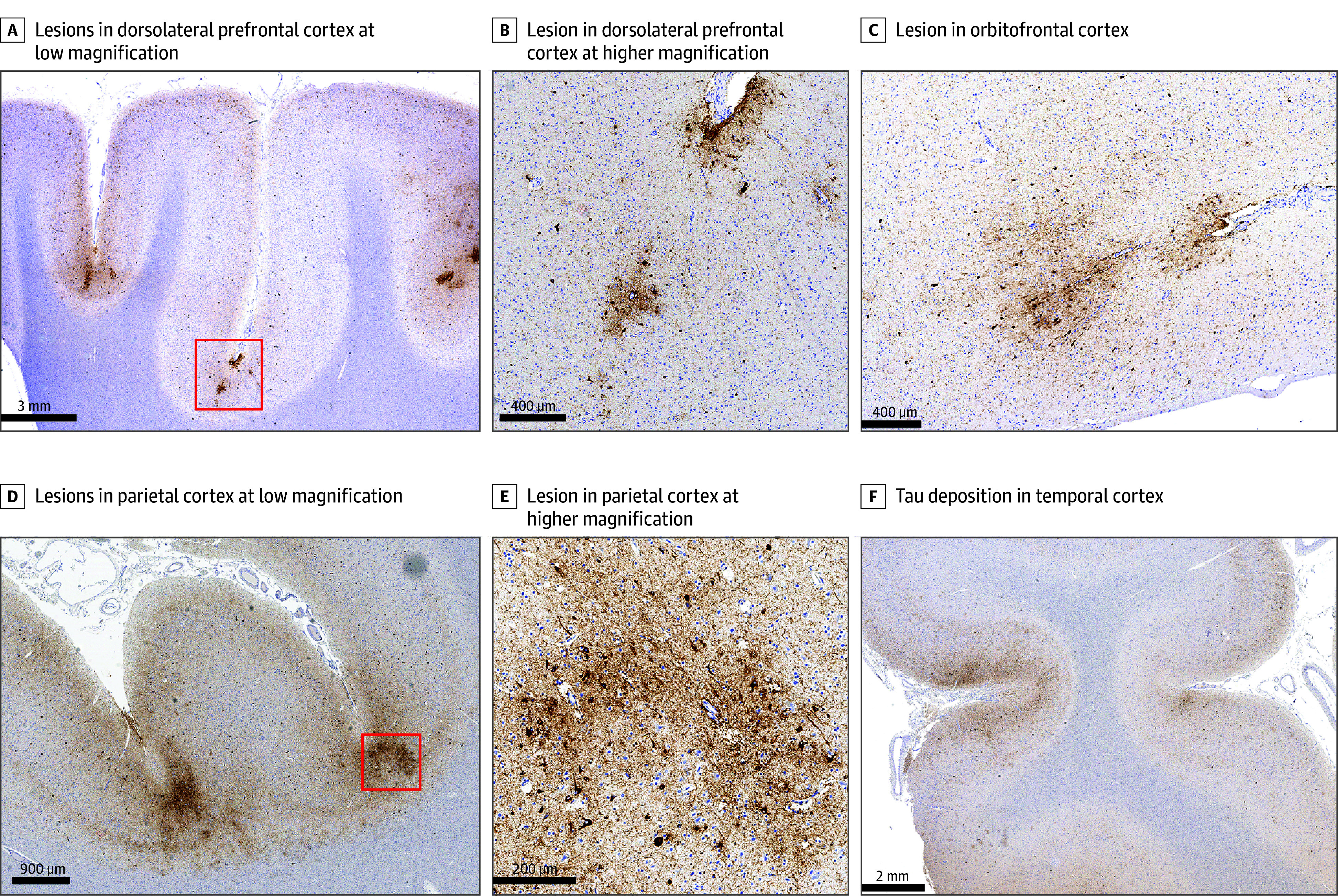
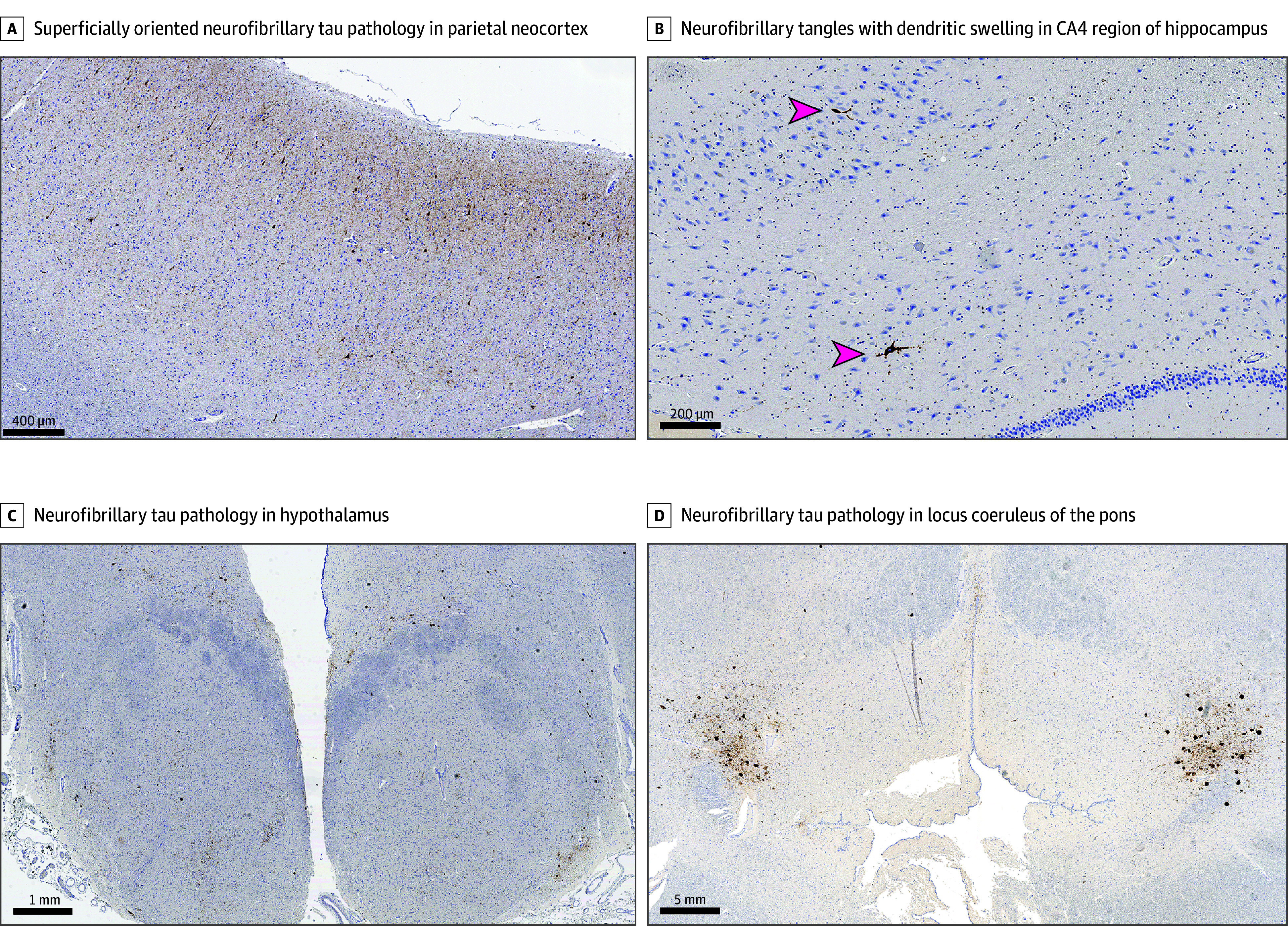
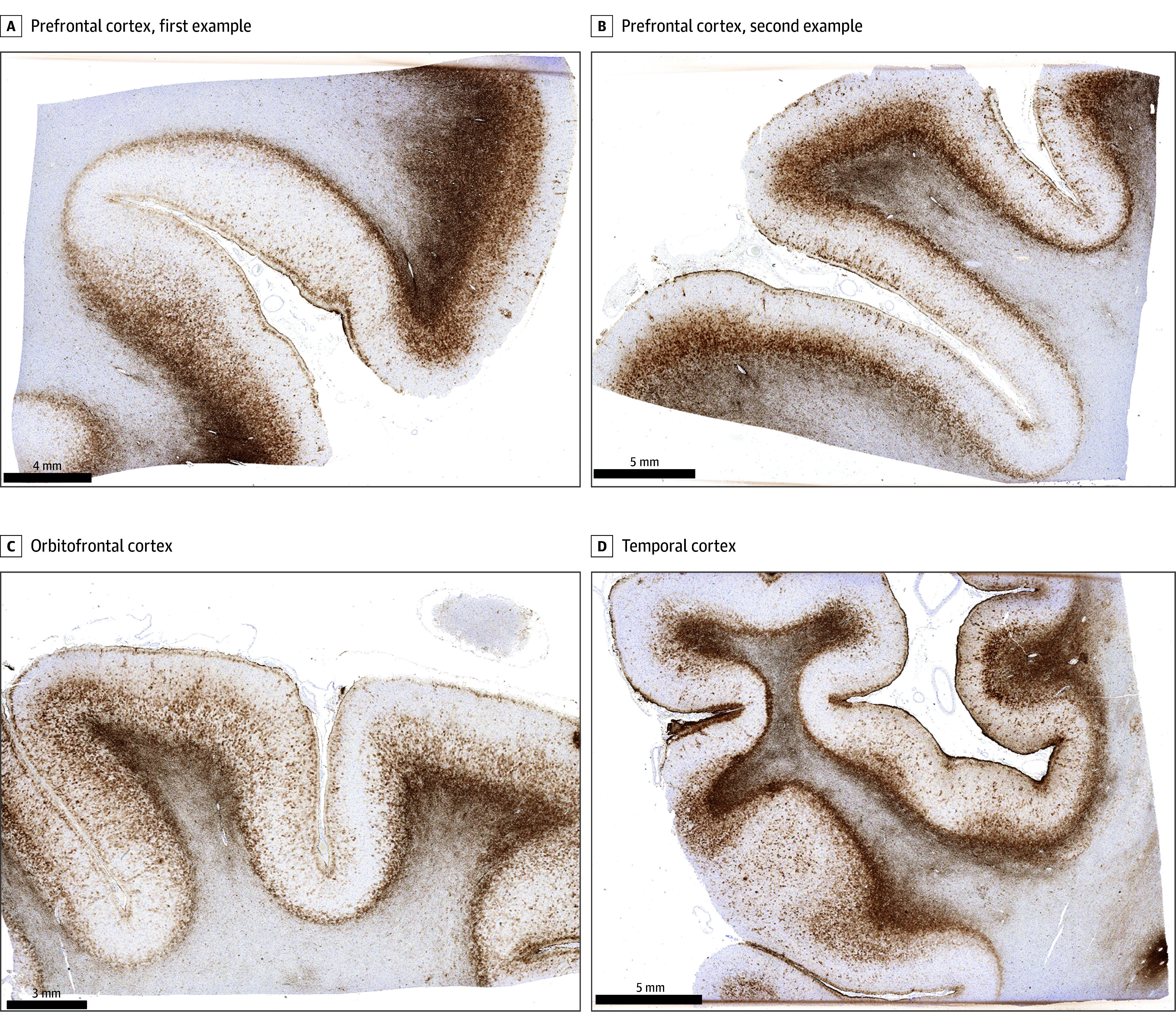
Results: The case specimen was from a male 44.9-year-old who served a 12.2-year SWCC career from ages 28.9 to 41.1 years. Following his last deployment, he experienced posttraumatic stress disorder, alcohol misuse, migraines, and impaired cognition. He died by suicide. Neuropathological evaluation revealed severe CTE (high CTE according to NINDS-NIBIB consensus criteria; McKee Stage IV of IV), with extensive pathology involving numerous brain regions. Molecular genetic testing for neurodegenerative disease risk was negative.

Conclusions and relevance: In this neuropathological case study of a 44-year-old former SWCC, we found severe CTE. Given the severity of the pathology at a relatively young age, we consider that exposures of a SWCC career, including repetitive physical forces applied to the head from high-speed boat impacts with waves, may be sufficient to promote or otherwise contribute to CTE development.


Conflict of interest statement

Conflict of Interest Disclosures: None reported.

Figures

Figure 1.
Figure 1.. Severe, Multilobar Chronic Traumatic Encephalopathy (CTE) in a Naval Servicemember, AT8 Immunohistochemistry for Phosphorylated Tau Protein
A, Demonstrates sulcal depth tau lesions characteristic of CTE in contiguous sulci at low magnification (scale bar = 3 mm) in a section from the dorsolateral prefrontal cortex; red box corresponds to region magnified in B. B, Demonstrates a pathognomonic lesion for CTE from the dorsolateral prefrontal cortex (corresponding to the region of the red box in A) at higher magnification (scale bar = 400 μm), characterized particularly by perivascular neuronal tau accumulation at a cortical sulcal depth. C, Additional example of another CTE lesion from a sample of the orbitofrontal cortex (scale bar = 400 μm). D, Demonstrates CTE lesions in contiguous sulci in a sample from the parietal lobe at low magnification (scale bar = 900 μm), red box corresponds to the region magnified in E. E, Demonstrates an extensive perivascular neuronal tau accumulation at a sulcal depth in the parietal cortex (corresponding to the region of the red box in D) at higher magnification (scale bar = 200 μm). F, Demonstrates extensive generalized tau deposition in the temporal cortex, with some concentration at the sulcal depths, at low magnification (scale bar = 2 mm).
Figure 2.
Figure 2.. Extensive Tau Pathology Corresponding to Secondary Characteristics of Chronic Traumatic Encephalopathy (CTE), AT8 Immunohistochemistry for Phosphorylated Tau Protein
A, Beyond sulcal depth lesions which are pathognomonic for CTE, this case additionally demonstrated diffuse cortical neuronal tau deposition, particularly in superficial layers of the cortex, which was prominent in the frontal, temporal, and parietal cortices and more minimal in the occipital sampling. This pattern of superficial cortical tau spread is commonly observed in severe cases of CTE; photomicrograph of a section from the parietal neocortex (scale bar = 400 μm). B, Demonstrates prominent neurofibrillary tangles (arrows) with dendritic swellings in the CA4 region of the hippocampus (scale bar = 200 μm), known to be a common secondary characteristic of CTE. C, Demonstrates extensive neurofibrillary tau pathology in the hypothalamus, including the mammillary bodies, at low magnification (scale bar = 1 mm). D, Demonstrates extensive neurofibrillary tau pathology in the locus coeruleus of the pons, at low magnification (scale bar = 5 mm).
Figure 3.
Figure 3.. Microscopic Features of Interface Astroglial Scarring
A-D (scale bars = 4 mm, 5 mm, 3 mm, and 5 mm, respectively), glial fibrillary acidic protein immunohistochemistry demonstrating substantially increased labeling, corresponding to astroglial scarring, particularly at subpial and gray-white interfaces, in select sections of the dorsolateral prefrontal cortex (A, B), orbitofrontal cortex (C), and temporal cortex (D). This pathology has been most predominantly described in the setting of prior blast-type traumatic brain injury.

Similar articles

References

    1. McKee AC, Cantu RC, Nowinski CJ, et al. Chronic traumatic encephalopathy in athletes: progressive tauopathy after repetitive head injury. J Neuropathol Exp Neurol. 2009;68(7):709-735. doi: 10.1097/NEN.0b013e3181a9d503 - DOI - PMC - PubMed
    1. McKee AC, Stern RA, Nowinski CJ, et al. The spectrum of disease in chronic traumatic encephalopathy. Brain. 2013;136(Pt 1):43-64. doi: 10.1093/brain/aws307 - DOI - PMC - PubMed
    1. Priemer DS, Iacono D, Rhodes CH, Olsen CH, Perl DP. Chronic traumatic encephalopathy in the brains of military personnel. N Engl J Med. 2022;386(23):2169-2177. doi: 10.1056/NEJMoa2203199 - DOI - PubMed
    1. Roesch JR, Curley M, Hart B. Sudden impacts in Naval Special Warfare high-speed boats. Naval Surface Warfare Center report No. CSS/TR-94/38. September 1994. Accessed November 13, 2024. https://combatantcraftcrewman.org/wp-content/uploads/2021/12/Impact-Repo...
    1. Kearns SD. Analysis and mitigation of mechanical shock effects on high speed planing boats. Massachusetts Institute of Technology graduate thesis. September 2001. Accessed November 13, 2024. https://dspace.mit.edu/handle/1721.1/8235

Publication types