. 2025 Jun 26;20(6):e0326978.
doi: 10.1371/journal.pone.0326978. eCollection 2025.

The Hippo-YAP/β-catenin signaling axis coordinates odontogenic differentiation in dental pulp stem cells: Implications for dentin-pulp regeneration

Affiliations

The Hippo-YAP/β-catenin signaling axis coordinates odontogenic differentiation in dental pulp stem cells: Implications for dentin-pulp regeneration

Chang Chen et al. PLoS One. .

Abstract

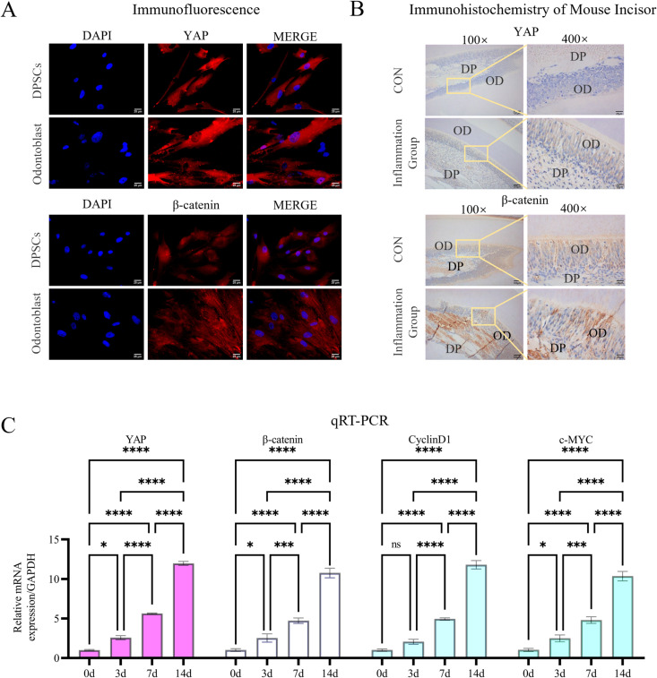
Objective: This study investigated the interplay between Hippo-YAP and β-catenin signaling in regulating odontogenic differentiation of human dental pulp stem cells (DPSCs) and explored its potential implications for dentin-pulp regeneration.

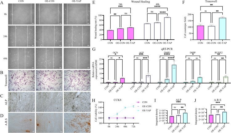
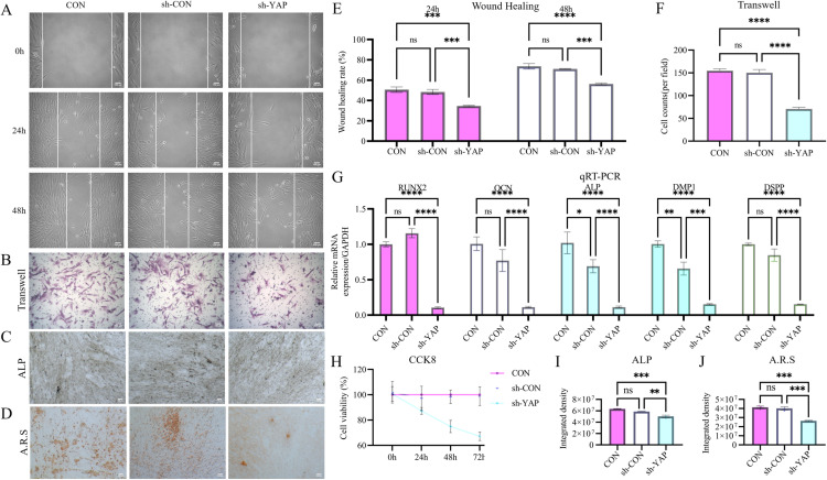
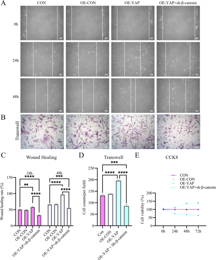
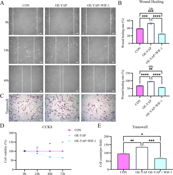
Methods: Using lentivirus-mediated YAP overexpression/silencing, β-catenin siRNA knockdown, and pharmacological Wnt inhibition (via WIF-1), we assessed DPSC proliferation, migration, mineralization, and molecular markers (via qRT-PCR, immunofluorescence). In vivo validation employed subcutaneous transplantation of DPSC-seeded scaffolds in immunocompromised mice.

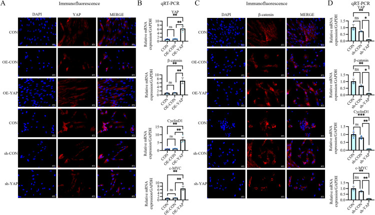
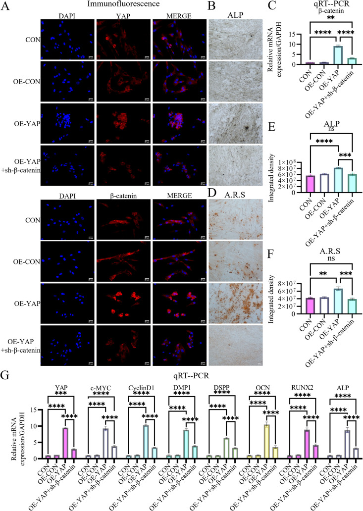
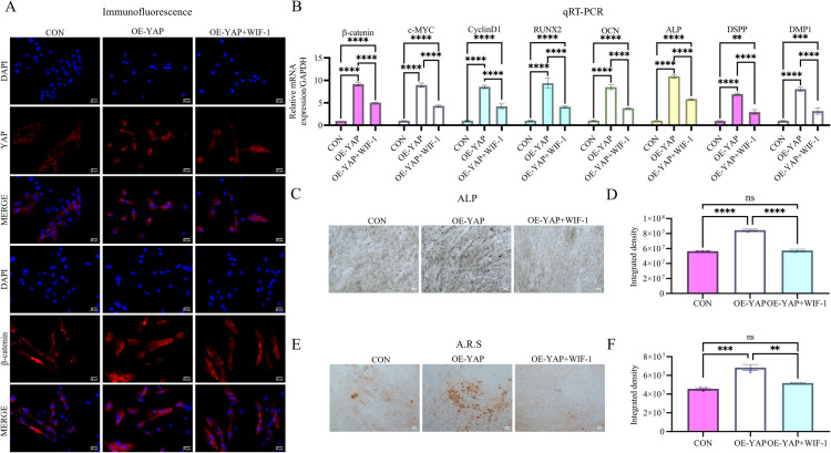
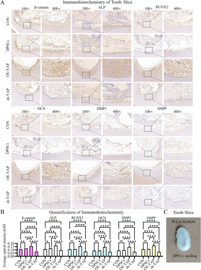
Results: YAP activation enhanced DPSC proliferation (1.44-fold), migration (1.39-fold), invasion (1.54-fold), and differentiation, as evidenced by elevated ALP activity (1.46-fold) and mineralization (1.36-fold). We observed transcriptional upregulation of odontogenic markers (RUNX2, DSPP, DMP1, OCN, ALP) and Wnt pathway components (β-catenin, Cyclin D1, c-Myc). Immunofluorescence revealed coordinated YAP and β-catenin expression patterns during differentiation. β-catenin silencing or Wnt inhibition abolished YAP-mediated functional enhancements and simultaneously suppressed YAP expression, partially confirming bidirectional regulation. In vivo, YAP-overexpressing DPSCs exhibited 1.27- to 1.62-fold induction of dentin-specific markers and β-catenin, whereas YAP silencing impaired these markers expression.

Conclusions: Our findings demonstrate that coordinated YAP and β-catenin signaling drives DPSC odontogenesis, with potential implications for dentin regeneration. Although reciprocal regulation is evident, direct molecular interactions require further validation.


Conflict of interest statement

The authors have declared that no competing interests exist.

Figures

Fig 1
Fig 1. Spatiotemporal activation dynamics of YAP and β-catenin during DPSC odontogenic differentiation.
(A) Immunofluorescence shows progressive increases in YAP and β-catenin protein levels in DPSCs over 14 days of odontogenic induction. Nuclei were stained with DAPI (blue). Scale bar: 25 µm. (B) Spatial immunohistochemical mapping in hydrogen peroxide-bleached murine incisors (mimicking inflammatory pulp conditions) demonstrates enriched YAP/β-catenin co-expression in odontoblast layers adjacent to predentin. Scale bar: 100 µm in100 × ; 25 µm in 400 × . (C) qRT-PCR quantification confirms progressive upregulation of YAP, β-catenin, and Wnt targets (Cyclin D1, c-Myc) during differentiation. Data: mean ± SD; *P < 0.05, ** P < 0.01, *** P < 0.001, **** P < 0.0001.
Fig 2
Fig 2. YAP overexpression potentiates DPSCs’ regenerative capacity.
(A, E) Scratch wound healing assay shows a 1.39-fold accelerated closure in YAP-overexpressing DPSCs (OE-YAP) vs. controls at 48 h. (B, F) Transwell invasion assay demonstrates a 1.54-fold increased invasion capacity in OE-YAP cells. (C, I) Alkaline phosphatase (ALP) staining and activity quantification reveal a 1.46-fold elevation in OE-YAP cells at day 14. (D, J) Alizarin Red staining (red nodules) and mineralization quantification show a 1.36-fold higher mineralization in OE-YAP cells. (G) qRT-PCR confirms that OE-YAP upregulates odontogenic markers (RUNX2, DSPP, DMP1, OCN, and ALP). (H) CCK-8 proliferation assay shows a 1.44-fold increase in OE-YAP cells at 72 h. Data: mean ± SD; *P < 0.05, ** P < 0.01, *** P < 0.001, **** P < 0.0001. Scale bar: 100 µm.
Fig 3
Fig 3. YAP knockdown suppresses DPSCs’ regenerative properties.
(A–J) Functional assays demonstrate that YAP silencing (sh-YAP) significantly attenuates key regenerative capacities compared to controls: (i) wound healing (A, E); (ii) Transwell invasion (B, F); (iii) ALP activity (C, I); (iv) mineralization (D, J); (v) proliferation (H). (G) qRT-PCR confirms that the sh-YAP group downregulates odontogenic markers. Data: mean ± SD; *P < 0.05, ** P < 0.01, *** P < 0.001, **** P < 0.0001. Scale bar: 100 µm.
Fig 4
Fig 4. YAP enhances β-Catenin and Wnt-related gene expression in odontogenic DPSCs.
(A) Lentiviral-mediated YAP overexpression (OE-YAP) and knockdown (sh-YAP) efficiency validated by immunofluorescence. (B, D) qRT-PCR shows OE-YAP upregulates Wnt pathway components (β-catenin, c-Myc, and Cyclin D1), while sh-YAP suppresses them. (C) Immunofluorescence reveals OE-YAP increases cytoplasmic β-catenin (red), whereas sh-YAP diminishes it. Data: mean ± SD; *P < 0.05, ** P < 0.01, *** P < 0.001, **** P < 0.0001. Scale bar: 25 µm.
Fig 5
Fig 5. β-catenin ablation reverts YAP-driven functional enhancement.
(A, C) The scratch wound closure assay: β-catenin siRNA reduces OE-YAP migration by 42.87% at 48 h (representative images in A, quantification in C). (B, D) The Transwell assay: β-catenin knockdown decreases OE-YAP invasion by 56.12% (representative images in B, quantification in D). (E) CCK-8 analysis shows that β-catenin siRNA reduces OE-YAP proliferative capacity by 54.97%. Data: mean ± SD; *P < 0.05, ** P < 0.01, *** P < 0.001, **** P < 0.0001 vs. control. Scale bar: 100 µm.
Fig 6
Fig 6. β-catenin ablation suppresses YAP expression and odontogenic capacity.
(A) Immunofluorescence demonstrates that β-catenin knockdown not only abolishes OE-YAP-induced β-catenin upregulation but also reduces YAP expression, indicating β-catenin-dependent stabilization of YAP. Scale bar: 25 µm. (C) qRT-PCR validates β-catenin siRNA knockdown efficacy. (B, E) ALP activity is decreased by 25.48% in β-catenin-silenced OE-YAP cells compared to controls. Scale bar: 100 µm. (D, F) Mineralization capacity is reduced by 41.52% following β-catenin ablation. Scale bar: 100 µm. (G) β-catenin siRNA downregulates YAP-induced markers and reciprocally suppresses YAP mRNA expression. Data: mean ± SD; *P < 0.05, ** P < 0.01, *** P < 0.001, **** P < 0.0001.
Fig 7
Fig 7. Pharmacological Wnt inhibition mimics β-catenin silencing in suppressing regenerative potential.
(A-D) Wnt inhibitory factor-1 (WIF-1) treatment phenocopies β-catenin siRNA effects, including wound closure reduction (AB), invasion capacity decrease (CE), and proliferation decline (D). Data: mean ± SD; *P < 0.05, ** P < 0.01, *** P < 0.001, **** P < 0.0001. Scale bar: 100 µm.
Fig 8
Fig 8. Pharmacological Wnt inhibition mimics β-catenin silencing in suppressing molecular expression and odontogenic differentiation.
(A) Immunofluorescence demonstrates diminished expression of both YAP and β-catenin in WIF-1-treated OE-YAP cells. Scale bar: 25 µm (B) qRT-PCR confirms WIF-1-mediated downregulation of β-catenin and its downstream targets c-Myc and Cyclin D1 in OE-YAP cells. (CD) WIF-1 treatment abolishes YAP-mediated enhancement of ALP activity and mineralization capacity. Scale bar: 100 µm. Data: mean ± SD; *P < 0.05, ** P < 0.01, *** P < 0.001, **** P < 0.0001.
Fig 9
Fig 9. In vivo validation of YAP-mediated dentin regeneration.
(A) Immunohistochemistry of implanted DPSCs grafts shows OE-YAP upregulates dentinogenic markers (DSPP, DMP1, RUNX2, OCN, and ALP) and β-catenin compared to controls. Scale bar: 100 µm (100×); 25 µm (400×). (B) Quantification confirms 1.36- to 1.62-fold induction of odontogenic markers and 1.27-fold β-catenin increase in OE-YAP grafts. (C) Schematic illustration of the tooth slice/scaffold design for ectopic transplantation. Data: mean ± SD; *P < 0.05, ** P < 0.01, *** P < 0.001, **** P < 0.0001.

Similar articles

References

    1. GBD 2021 Oral Disorders Collaborators. Trends in the global, regional, and national burden of oral conditions from 1990 to 2021: a systematic analysis for the Global Burden of Disease Study 2021. Lancet. 2025;405(10482):897–910. doi: 10.1016/S0140-6736(24)02811-3 - DOI - PubMed
    1. Sui BD, Jin Y. Theory and practice of mesenchymal condensation directing tooth development and regeneration. Zhonghua Kou Qiang Yi Xue Za Zhi. 2024;59(5):418–25. doi: 10.3760/cma.j.cn112144-20240131-00060 - DOI - PubMed
    1. Shao C, Jin B, Mu Z, Lu H, Zhao Y, Wu Z, et al. Repair of tooth enamel by a biomimetic mineralization frontier ensuring epitaxial growth. Sci Adv. 2019;5(8):eaaw9569. doi: 10.1126/sciadv.aaw9569 - DOI - PMC - PubMed
    1. Wang Y, Mao J, Wang Y, Wang R, Jiang N, Hu X, et al. Odontogenic exosomes simulating the developmental microenvironment promote complete regeneration of pulp-dentin complex in vivo. J Adv Res. 2025:S2090-1232(24)00626-X. doi: 10.1016/j.jare.2024.12.048 - DOI - PubMed
    1. Jiang L, Li J, Zhang C, Shang Y, Lin J. YAP‑mediated crosstalk between the Wnt and Hippo signaling pathways (Review). Mol Med Rep. 2020;22(5):4101–6. doi: 10.3892/mmr.2020.11529 - DOI - PubMed

MeSH terms