. 2025 Jun 26;21(6):e1013255.
doi: 10.1371/journal.ppat.1013255. eCollection 2025 Jun.

Discovery of functional factorless internal ribosome entry site-like structures through virome mining

Affiliations

Discovery of functional factorless internal ribosome entry site-like structures through virome mining

Subash Chapagain et al. PLoS Pathog. .

Abstract

All viruses must co-opt the host translational machinery for viral protein synthesis. The dicistrovirus intergenic region internal ribosome entry site (IGR-IRES) utilizes the most streamlined translation mechanism by adopting a triple pseudoknot structure that directly recruits and binds within the intersubunit space of the ribosome and initiates translation from a non-AUG codon. The origin of this unprecedented mechanism is not known. Using a bioinformatics pipeline to examine the diversity and function of IRESs across RNA viromes, we searched for IRES-like RNA structures using RNA covariance models for multiple IRES sub-types, and tested functional IRES by using a dual-fluorescent lentiviral library reporter screen. We identified over >4,700 dicistro-like genomes with ~32% containing putative IRES structures, including novel viral genome arrangements with multiple IRESs and IRESs embedded within open-reading frames (ORFs). Predicted IRESs bound directly to purified ribosomes and supported internal ribosome entry activity in vitro and in vivo. Moreover, internal IRESs embedded within an ORF of monocistronic genomes were functional and operated simultaneously to produce the downstream ORF. We also identified IRES-like structures within non-dicistrovirus viral genomes, including in the families Tombusviridae and Narnaviridae that bound to ribosomes directly and a subset can direct internal ribosome entry. This study provides a framework to map the origin of factorless IRES mechanisms and study the diverse viral strategies utilizing RNA-based mechanisms.


Conflict of interest statement

The authors have declared that no competing interests exist.

Figures
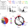
Fig 1
Fig 1. Global discovery of Type 6 internal ribosome entry sites.
(A) Schematic of the bioinformatic pipeline to identify structures that are Type 6 IRES-like. Dicistrovirus genomes were selected from metatranscriptomes and the earth’s virome project (Serratus.io). Transcripts containing an RdRP sequence with >50% amino acid identity from ICTV-classified dicistroviruses were selected using Palsmscan. Genomes were clustered at 98% nucleotide identity to eliminate redundant sequences, followed by selecting >2 kb genomes that contain at least 1 ORF > 1 kb in length (9,151 genomes, S1 Data). Type 6 RNA covariance models based on currently described IRES subtypes were generated and used to scan the final dataset using INFERNAL. (B) Secondary structure schematics of Type 6 IRESs. (C) Distribution of Type 6 IRES-like structures in dicistrovirus genomes by genome size. Total genomes (light grey bars) and genomes containing IRESs (black bars) are shown. (D) Pie chart of the distribution of Type 6 IRES subtypes identified by INFERNAL. Genomes identified by multiple covariance models in the INFERNAL search are shown (S1 Data) (E) Distribution of Dicistrovirus genomes based on their genomic length. Shown in bar graphs is the number of genomes with or without IRES-like structures predicted by INFERNAL (F) Distribution of Type 6 IRES subtypes in an RdRP-based phylogenetic tree. RdRP amino acid sequences from genomes >6 kb were aligned using MUSCLE and a maximum likelihood tree was built with 1000 pseudo bootstrap branch support. IRES subtypes are identified with lines on the outside circle, colour-coded based on subtype. Sequences with multiple IRESs are shown with an extra stacked line. Genomes with IRESs that were used to build covariance models are shown on the outer ring. Virus names are specified for known dicistroviruses from the Cripavirus, Aparavirus, and Triatovirus genera (ICTV). Genomes and IRESs used to build the phylogenetic tree are listed in S1 and S2 Data.
Fig 2
Fig 2. Consensus models of Type 6 IRES.
Secondary structure models of indicated Type 6 IRESs are shown with colour-coded heat map of nucleotides representing percent conservation from alignment of the top 50% scoring sequences for each subtype based on INFERNAL output (S2 Data). The range of base pairing are also denoted and the number of sequences used for alignment of each sub-type are indicated. Pie charts show the percentages of the predicted codon adjacent to the pseudoknot I (PKI) IRES sequences. The resulting multiple sequence alignments were manually curated to remove gaps.
Fig 3
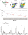
Fig 3. Functional analysis of IRES-like RNAs.
(A) Schematic of IRES screening pipeline in mammalian cells. A library of IRESs were cloned within the intergenic region of a lentivirus eGFP-mRuby3 dual reporter construct. HEK293 cells were transduced with lentivirus and fluorescent cells were analyzed by FACS analysis and sorted. Cells were either untreated or treated with Thapsigargin (1 μM) for 18 hours. (B) FACS plots of cells eGFP (y-axis) and mRuby3 (x-axis) fluorescence. The percent of mRuby3 positive cells are shown under specific cell treatments. IRESs identified in untreated and Thapsigargin treated cells are listed in S3 Data. (C) Sequencing read counts for IRESs in mRuby3 positive cells transduced with lentiviral reporter library in untreated (red) and Thapsigargin treated (blue) conditions. The counts are ranked from highest to lowest for IRESs enriched in untreated conditions. All raw read counts for unfiltered and filtered IRESs are in S3 Data. (D) In vitro translation. Select Type 6a and 6b IRESs were tested in (left graph) rabbit reticulocyte lysates (1 hour, 37°C) or (right graph) in Sf21 insect lysates (2 hours, 30°C) using in vitro transcribed dual luciferase reporter RNAs. IRESs selected are listed in S4 Data. Shown are averages ± s.d. from at least three independent experiments of FLuc/RLuc luciferase activity ratios normalized to that of bicistronic RNAs containing the wild-type (WT) CrPV IGR IRES. mPKI represents mutant CrPV IRES containing mutations that disrupt PKI base pairing. INFERNAL scores are shown below the indicated IRESs. Shown are averages ± s.d. from at least three independent experiments.
Fig 4
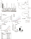
Fig 4. Integrity of IRES structure for translation and ribosome binding.
(A) Integrity of PKI for IRES function. Bicistronic luciferase reporter RNAs containing select IRESs containing either mutations that disrupt PKI base pairing (mPKI) or compensatory mutations that restore PKI base pairing (cPKI) were incubated in RRL for 1 hour, 37°C. Shown are averages of FLuc/RLuc luciferase activity ratios normalized to that of bicistronic RNAs containing the wild-type (WT) CrPV IGR IRES. mPKI represents mutant CrPV IRES containing mutations that disrupt PKI base pairing. (B) Secondary structure model of the Type 6b-2 IGR IRES. Mutations that disrupt the PKI base pairing and compensatory mutations that restore the base pairing are indicated in rectangular boxes. (C) 80S ribosome binding. Filter binding assay of purified, salt washed human 40S and 60S incubated with select [32P]-labeled RNAs (0.5 nM) and the fraction of IRES:ribosome complexes were measured by phosphorimager analysis. mPKII/PKIII and mPKI/mPKII/mPKIII represent mutant IGR IRESs whereby PKI, PKII and PKIII base pairing are disrupted. (D) Apparent dissociation constant (KD) values of 80S:IRES complexes. (E) Relative luciferase activity of Type 6a IRES subtype in S2 cells. S2 cells were transfected with reporter RNAs containing the indicated wild-type 6a-11 IRES or CrPV IRES (WT), mPKI (mutation in IRES that disrupts PKI base pairing). Shown are averages of Firefly:Renilla (Fluc:RLuc) luciferase activities at 6 hours post-transfection. All data are averages ± s.d. from at least three independent experiments.
Fig 5
Fig 5. Genome architectures of dicistrovirus-like genomes.
Genome organization of dicistrovirus genomes was obtained by predicting ORFs using ORffinder, RdRps with Palmscan, and IRESs with INFERNAL. ORF2 > 1 kb in length were predicted using any possible non-stop codon as a start codon. The number of genomes within specific sub-types and the total numbers are shown to the right. Genomes are categorized with predicted IRESs located within the 5′ UTR or a single long ORF, or genomes with multiple predicted IRESs. Genomes containing predicted IRES with more than two ORFs are shown. Predicated helicase (Hel) and 3C-like protease (Pro) and RNA-dependent RNA polymerase (RdRP) are shown. Genomes are listed in S5 Data.
Fig 6
Fig 6. Functional analysis of IRES within an atypical dicistrovirus genome.
(A) Schematic of the Type 6a-2 IRES (type 6a-2) within the atypical dicistrovirus genome. Predicted secondary structure model of the Type 6a-2 IRES. (B) Schematic of reporter RNAs mirroring their locations in atypical dicistrovirus genome. “RS” denotes a stop codon at the end of the Renilla luciferase ORF (stop codon is represented by a red octagon). “HP” denotes a strong hairpin stem-loop inserted within the 5′ UTR. The predicted translated protein masses are shown above. (C) SDS-PAGE analysis of in vitro translation reactions (top) and quantification of proteins (bottom). In vitro-transcribed bicistronic reporter RNA (600 ng) containing the indicated wild-type or mutant IRESs were incubated in RRL (1 hour, 37°C) with [35S]-methionine. (D) IRES-mediated translation in mock and CrPV-infected (MOI 10) S2 cells. S2 cells were transfected with reporter RNAs containing the indicated wild-type 6a-2 IRES or CrPV IRES (WT), mPKI (mutation in IRES that disrupts PKI base pairing), RS (Renilla with a stop codon) or HP (hairpin within the 5′ UTR). Shown are averages of Firefly (FLuc) and Renilla (RLuc) luciferase activities ± s.d. from at least three independent experiments at 6 hours post-transfection.
Fig 7
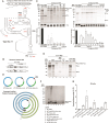
Fig 7. Translation of embedded contiguous IRESs.
(A) Genomic location (top schematic) of a representative embedded IRES (type 6a-11) and its predicted secondary structure (bottom). P2A – denotes 2A peptide from porcine tescovirus-1. Scar – denotes sequence left over after RNA circularization. (B) Schematic of the luciferase reporter RNAs containing the IRES embedded within the ORF mirroring the location of the IRES in the native monocistronic genome. Predicted MW of RLuc (36.0 kDa), FLuc (61.7 kDa) and fusion RLuc-IRES-FLuc (106 kDa) are shown. Stop codon is indicated as a red octagon. (C) Translation of luciferase reporters containing select embedded IRESs (listed in S4 Data). In vitro transcribed bicistronic reporter RNAs containing the indicated IRESs were incubated in RRL (1 hour, 37°C) with [35S]-methionine. Reactions were loaded on 12% SDS-PAGE gels and imaged by phosphorimager analysis. Representative gels (top panel) and quantification of relative Firefly luciferase band intensities (bottom graphs) are shown. (D) Mutational analysis of Type 6a-11 IRES. Representative SDS-PAGE gel of [35S]-methionine in vitro translation reactions of reporter RNAs in RRL. mPKI-III denotes mutations that disrupt PKI, PKII and PKIII base pairing, m4SL denotes mutations within SLIV, SLV and L1.1 collectively. “Stop ORF1” denotes a luciferase reporter wherein a stop codon is inserted at the end of Renilla luciferase as shown in (B). (E) Schematic of circular RNAs and predicted circRNA protein products. (F) Representative SDS-PAGE gel of in vitro translation (RRL) reactions containing [35S]-methionine and the indicated circRNA reporter. “SubP2A” denotes P2A sequence lacking the N-terminal GSG. 3mPK denotes mutations that disrupts all three pseudoknots within the IRES. (G) Translational activity of IRES circRNA in S2 cells. S2 cells were transfected with circular RNAs containing the embedded IRES. At 8 hours post-transfection, cells were harvested and lysed and luciferase activities were measured. Shown are averages ± s.d. from at least three independent experiments.
Fig 8
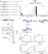
Fig 8. Functional IGR IRES in non-dicistrovirus viral genomes.
(A) Schematic of non-dicistrovirus genomes and the location of predicted Type 6 IRES. (B) Non-dicistrovirus-like IRES translation. In vitro transcribed dual luciferase reporter RNAs containing the indicated non-dicistrovirus predicted IRESs within the intergenic region were incubated in RRL (1 hour, 37°C) or Sf21 extracts (2 hours, 30°C). Luciferase activities were measured and the ratio of FLuc/RLuc was calculated and normalized to that of CrPV IRES containing reporter RNAs. (C) Genome organization of Tombusvirus-like non-DV1 genome with a predicted IGR IRES (top) and secondary structure model of the putative IRES (bottom). Mutations used for subsequent binding experiments are denoted in the boxes. (D) Predicted non-DV1 RNA structure model from SAXS data (light blue) overlapped with atomistic structure, represented in ribbons. (E) Sucrose gradient centrifugation analysis (top) showing percent total radioactive counts (y-axis) in fractions (top to bottom) of reactions containing [32P] labeled IRES incubated with purified salt-washed 40S and 60S. Wild-type (black lines) and mutant (grey lines) of CrPV IRES and non-DV-1 RNA are shown. Mutant IRES denotes mutations that disrupt both PKII and PKIII base pairing. Filter binding assays (bottom) of purified human 80S with indicated wild-type and mutant IRES. 32P labeled IRESs (0.5 nM) were incubated with increasing concentrations of purified, salt-washed human 40S and 60S subunits and the fraction of IRES:ribosome complexes were measured by phosphorimager analysis. Shown are averages ± s.d. from at least three independent experiments.

Similar articles

References

    1. Jan E, Mohr I, Walsh D. A Cap-to-Tail Guide to mRNA Translation Strategies in Virus-Infected Cells. Annu Rev Virol. 2016;3(1):283–307. doi: 10.1146/annurev-virology-100114-055014 - DOI - PubMed
    1. Mailliot J, Martin F. Viral internal ribosomal entry sites: four classes for one goal. Wiley Interdiscip Rev RNA. 2018;9(2). doi: 10.1002/wrna.1458 - DOI - PubMed
    1. Kwan T, Thompson SR. Noncanonical Translation Initiation in Eukaryotes. Cold Spring Harb Perspect Biol. 2019/04/01 ed. 4AD;11. doi: 10.1101/cshperspect.a032672 - DOI - PMC - PubMed
    1. Jang SK, Kräusslich HG, Nicklin MJ, Duke GM, Palmenberg AC, Wimmer E. A segment of the 5’ nontranslated region of encephalomyocarditis virus RNA directs internal entry of ribosomes during in vitro translation. J Virol. 1988;62(8):2636–43. doi: 10.1128/JVI.62.8.2636-2643.1988 - DOI - PMC - PubMed
    1. Jaafar ZA, Kieft JS. Viral RNA structure-based strategies to manipulate translation. Nat Rev Microbiol. 2019;17(2):110–23. doi: 10.1038/s41579-018-0117-x - DOI - PMC - PubMed

LinkOut - more resources