. 2025 Jul 9;482(14):969-988.
doi: 10.1042/BCJ20253107.

Structure and activity of the essential UCH family deubiquitinase DUB16 from Leishmania donovani

Affiliations

Structure and activity of the essential UCH family deubiquitinase DUB16 from Leishmania donovani

James A Brannigan et al. Biochem J. .

Abstract

In Leishmania parasites, as for their hosts, the ubiquitin (Ub) proteasome system is important for cell viability. As part of a systematic gene deletion study, it was discovered that four cysteine protease-type deubiquitinases (DUBs) are essential for parasite survival in the promastigote stage, including DUB16. Here, we have purified and characterised recombinant DUB16 from Leishmania donovani, which belongs to the Ub C-terminal hydrolase (UCH) family. DUB16 efficiently hydrolyses C-terminal aminocoumarin and rhodamine conjugates of Ub consistent with proposed cellular roles of UCH-type DUBs in regenerating free monomeric Ub from small molecule Ub adducts arising from adventitious metabolic processes. The crystal structure of DUB16 reveals a typical UCH-type DUB fold, and a relatively short and disordered cross-over loop that appears to restrict access to the catalytic cysteine. At close to stoichiometric enzyme to substrate ratios, DUB16 exhibits DUB activity towards diubiquitins linked through isopeptide bonds between Lys11, Lys48 or Lys63 residues of the proximal Ub and the C-terminus of the distal Ub. With 100-1000-fold higher turnover rates, DUB16 cleaves the ubiquitin-ribosomal L40 fusion protein to give the mature products. A DUB-targeting cysteine-reactive cyanopyrrolidine compound, IMP-1710, inhibits DUB16 activity. IMP-1710 was shown in promastigote cell viability assays to have parasite killing activity with EC50 values of 1-2 μM, comparable with the anti-leishmanial drug, miltefosine. L. mexicana parasites engineered to overproduce DUB16 showed a modest increase in resistance to IMP-1710, providing evidence that IMP-1710 inhibits DUB16 in vivo. While it is highly likely that IMP-1710 has additional targets, these results suggest that DUB16 is a potential target for the development of new anti-leishmanial compounds.

Keywords: UCH family; crystal structure; deubiquitinase; leishmania; substrate specificity.


Conflict of interest statement

The authors declare that there are no competing interests associated with this manuscript.

Figures

Figure 1
Figure 1. DUB16 acts on peptide and Ub conjugates.
Plots of initial velocity versus substrate concentration for DUB16 cleavage of Z-RLRGG-AMC (A) and Ub-RhoGly (B). (C) Coomassie stained SDS-polyacrylamide gel following electrophoresis to resolve DUB16, Ub-propargylamide (Ubi-PA) and their reaction product following mixing and co-incubation with increasing amounts of Ubi-PA to give molar ratios of 0.5, 1, 2 and 4. M, molecular weight markers; D, 3 μg DUB16; U, 2 μg ubiquitin.
Figure 2
Figure 2. Crystal structure of DUB16.
(A) Plot of A280 (blue line) versus elution time in a SEC-MALLS experiment performed for LdDUB16. The major peak at 23 minutes constitutes >90 % of the sample loaded and is associated with a molecular mass of 25 kDa (orange line) calculated from the light scattering and refractive index measurements. (B) Ribbon tracing of the backbone structure of DUB16. The chain is colour-ramped from the N-terminus (blue) to the C-terminus (pink). The secondary structure elements are labelled and the active site residues are shown in cylinder format. (C) The structure of DUB16 displayed as an electrostatic surface rendering (red, negative, blue positive). The view is essentially orthogonal to that in (B) looking down into the active site groove. The atoms of the catalytic cysteine 94 residue are shown as yellow spheres emphasising the recessed location of this residue. (D) The active site residues in the structure of DUB16 superimposed on the active site of the human UCHL3-UbVME complex following superposition of the main chain atoms of the A subunits in the two structures. The structures are distinguished by the green and grey carbon atoms respectively. The UbVME moiety has been omitted from the UCHL3 structure for clarity (E) Superposition of DUB16 (ice blue) and the human UCHL3-UbVME complex (UCHL3, grey: UbVME, gold). The chains are shown as ribbons except for the C-terminal residues of UbVME which are shown as cylinders coloured by atom type with carbons in gold, nitrogens in blue and oxygens in red. The side chains of the active cysteines of the DUBs are shown as cylinders with the carbons in green and the sulphurs in yellow. Elements of the DUB16 secondary structure which partially clash with the UbVME moiety, including the α8−β3 crossover loop are labelled. (F) Alignment of the sequences of the deubiquitinase domains of the UCH type DUBs prepared in the program TCoffee and displayed in the program ESPRIPT in the context of the secondary structure elements of DUB16. Invariant residues are highlighted by a red background – conserved residues by blue boxes. The black bar below the sequence spans the residues of the crossover loop, the triangles denote the active site residues. The asterisk above the sequence indicates alternate conformations of the residue.
Figure 3
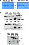
Figure 3. DUB16 displays linkage specificity in the cleavage of diubiquitin substrates.
Deubiquitinase and diubiquitin substrates with the indicated linkages were co-incubated for the indicated time in minutes prior to quenching with the addition of SDS-containing sample buffer and resolution of the products by 12 % polyacrylamide gel electrophoresis. In (A), the gel was stained with Coomassie blue. In (B), following Western transfer and probing with anti-ubiquitin antibody, the blots were visualised with the ChemiDoc system (BioRad). Mouse anti-DUB16 antibody [37] was used to immunoblot the enzyme as a loading control. (C) Cleavage of tri- and tetraubiquitins monitored as in B.
Figure 4
Figure 4. DUB16 cleaves the Ubiquitin-L40 precursor protein to produce monomeric ubiquitin.
(A) Ribbon rendering of the AlphaFold2 model AF_E9BMW6. A zinc metal is shown as a white sphere, with its surrounding cysteines and residues in the linker region shown as cylinders. (B) Time course of Ub-L40 cleavage by DUB16. 10 μg of Ub-L40 substrate was incubated with 30 ng of DUB16 at 24°C and reaction aliquots were removed over the time range 0–32 minutes and quenched in SDS PAGE sample buffer. The reaction products were resolved by denaturing 17.5 % polyacrylamide gel electrophoresis and visualised by staining with Coomassie blue dye. The disappearance of the Ub-L40 band of apparent Mr = 16 kDa is accompanied by the concomitant appearance of the two higher mobility species, L40 and Ub. D, DUB16 (300 ng), M, markers, U, ubiquitin (3.5 μg).
Figure 5
Figure 5. In vitro inhibition of DUB16 activity.
(A) Structures of the cyanopyrrolidine IMP-1711 and its derivative IMP-1710 bearing an alkyne tag. (B) Coomassie stained SDS-PAGE showing inhibition of Ub-40 cleavage by IMP-1710; M, Mwt markers, S, Ub-L40 substrate, +/-I corresponds to DUB16 preincubated (30 min) in the presence and absence of a 40-fold excess of IMP-1710 before addition to the Ub-L40 substrate and further incubation at room temperature for 30 min, U, 2 μg ubiquitin; E, 0.5 μg DUB16. (C) Differential scanning fluorimetry profile of 12.5 μM DUB16 in the presence and absence of 25 μM IMP-1711.
Figure 6
Figure 6. In vivo profiling of inhibitor activity.
(A) DUB activity-based probe assay. Two populations (labelled as [DUB16] A & B) of L. mexicana M379 promastigotes were transfected with the pNUS::DUB16 episomal expression vector and selected with G418. Cell lysates were incubated with the HA-Ubi-PA activity-based probe and resolved by SDS-PAGE. Total protein in the polyacrylamide gel was visualised with the BioRad stain free TGX system prior to transfer to a PVDF membrane. Western blotting with an anti-HA primary antibody and visualising with an HRP secondary antibody shows upregulation of DUB16 activity in the transfected populations. In the HA-Ubi-PA blot, the lower band (indicated by the solid arrowhead) represents DUB16, the upper band (indicated by the open arrowhead) is likely to be DUB17 [10]. Oligopeptidase B (OPB) is used as a loading control for the blot. (B) Bar chart representing the mean anti-HA signal corresponding to DUB16 activity, quantified using BioRad ImageLab. Error bars denote the standard deviation. (C) Does response curves showing the activity of IMP-1710 against promastigote forms of L. mexicana M379 and the derived strains overexpressing DUB16 using a cell viability assay. The EC50 values are given in Table 2.

References

    1. Burza S., Croft S.L., Boelaert M Leishmaniasis. Lancet. 2018;392:951–970. doi: 10.1016/S0140-6736(18)31204-2. - DOI - PubMed
    1. Mann S., Frasca K., Scherrer S., Henao-Martínez A.F., Newman S., Ramanan P., et al. A review of leishmaniasis: current knowledge and future directions. Curr. Trop. Med. Rep. 2021;8:121–132. doi: 10.1007/s40475-021-00232-7. - DOI - PMC - PubMed
    1. Croft S.L., Coombs G.H Leishmaniasis--current chemotherapy and recent advances in the search for novel drugs. Trends Parasitol. 2003;19:502–508. doi: 10.1016/j.pt.2003.09.008. - DOI - PubMed
    1. Brannigan J.A., Wilkinson A.J Drug discovery in leishmaniasis using protein lipidation as a target. Biophys. Rev. 2021;13:1139–1146. doi: 10.1007/s12551-021-00855-0. - DOI - PMC - PubMed
    1. Saldivia M., Lima A.P.C.A., Mottram J.C A promising pipeline of preclinical drug candidates for leishmaniasis and chronic chagas’ disease. Trends Parasitol. 2024;40:211–213. doi: 10.1016/j.pt.2024.02.002. - DOI - PubMed

MeSH terms