. 2025 Jun 26;16(1):5372.
doi: 10.1038/s41467-025-60332-0.

Daily electric field treatment improves functional outcomes after thoracic contusion spinal cord injury in rats

Affiliations

Daily electric field treatment improves functional outcomes after thoracic contusion spinal cord injury in rats

Bruce Harland et al. Nat Commun. .

Abstract

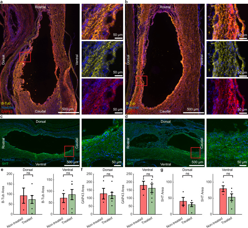
Spinal cord injury (SCI) can cause permanent loss of sensory, motor, and autonomic functions, with limited therapeutic options available. Low-frequency electric fields with changing polarity have shown promise in promoting axon regeneration and improving outcomes. However, the metal electrodes used previously were prone to corrosion, and their epidural placement limited the penetration of the electric field into the spinal cord. Here, we demonstrate that a thin-film implant with supercapacitive electrodes placed under the dura mater can safely and effectively deliver electric field treatment in rats with thoracic SCI. Subdural stimulation enhanced hind limb function and touch sensitivity compared to controls, without inducing a neuroinflammatory response in the spinal cord. While axon density around the lesion site remained unchanged after 12 weeks, in vivo monitoring and electrochemical testing of electrodes indicated that treatment was administered throughout the study. These results highlight the promise of electric field treatment as a viable therapeutic strategy for achieving long-term functional recovery in SCI.


Conflict of interest statement

Competing interests: The authors declare no competing interests.

Figures

Fig. 1
Fig. 1. Daily EF treatment improves hind limb motor function and touch sensitivity after SCI.
a Bioelectronic implant with stimulation electrodes. b Implant was inserted below T10–T12 in treated, non-treated, and no-injury rats. c A 175 kilodyne spinal cord impact at T11 showed no difference between groups. d Starting the day after surgery, treated rats received 2 Hz biphasic EF stimulation (1 h/day) via active and counter electrodes flanking the injury. e Treated rats initially recovered slower but surpassed non-treated rats in open-field motor scores from week 4 onward (green asterisks). f Treated rats showed faster hind paw withdrawal to ramping filament force at week 1 and weeks 3–12 compared to non-treated rats (green asterisks) and at week 4 and 9 compared to no-injury rats (blue asterisks). g Treated and non-treated rats showed similar foot misplacements on a 1-m ladder. h Treated rats had more slips on the ladder at weeks 3, 4, and 9 compared to non-treated (green asterisks). *P < 0.05, **P < 0.01, ***P < 0.001, ****P < 0.0001. Group means are shown by thick lines ± SEM; thin lines represent individual animals from treated and non-treated groups in (e, g, h). c, e Treated n = 8, Non-treated n = 10, No injury n = 10; fh Treated n = 8, Non-treated n = 8, No injury n = 10. c Two-way unpaired t-test, p = 0.94. eh Two-way repeated measures ANOVA - Tukey’s post hoc; e post hoc treated vs non-treated effects: w1: p = 0.026, w4: p = 0.039, w5: p = 0.014, w6: p = 0.003, w7: p = 0.0002, w8: p < 0.0001, w9: p = 0.0013, w10: p = 0.0007, w11: p = 0.010, w12: p = 0.0002; f post hoc treated vs non-treated effects: w1: p = 0.047, w3: p = 0.027, w4: p = 0.0023, w5: p = 0.0066, w6: p = 0.0061, w7: p = 0.016, w8: p = 0.0092, w9: p = 0.0016, w10: p = 0.0063, w11: p = 0.017, w12: p = 0.0008, post hoc treated vs no injury effects: w4: p = 0.02, w9: p = 0.037; g post hoc treated vs non-treated effects: p’s > 0.17; h post hoc treated vs non-treated effects: w3: p = 0.011, w4: p = 0.011, w9: p = 0.017.
Fig. 2
Fig. 2. Subdural administration of daily EF treatment did not result in a neuroinflammatory response.
Representative sagittal images are shown of immunolabelling of IBA1 and GFAP for a non-treated and b treated rats. c Dorsal and Ventral regions of tissue were analyzed. d There was no significant difference in microglial/macrophage activity between the treated and non-treated groups in the dorsal and ventral regions of the spinal cord. e No difference in astrocyte activity was detected between groups. Data are presented as mean values ± SEM. d, e treated n = 6, non-treated n = 3; two-way unpaired t-tests. d Dorsal: p = 0.30, ventral: p = 0.32; e dorsal: p = 0.87, ventral: p = 0.24.
Fig. 3
Fig. 3. There were no changes in axon density or serotonergic expression in rats given daily EF treatment compared with non-treated controls.
a Representative sagittal images from the epicenter of the lesion with β-tub and GAP43 immunolabelling from a non-treated and b treated rats. Representative sagittal images from the epicenter of the lesion with 5HT immunolabelling from c non-treated and d treated rats. Similar expression of both e β-tub (overall axon density) and f GAP43 (axonal regeneration) were observed in both groups, either in the dorsal or ventral regions of the spinal cord. g Similar levels of serotonergic (5HT) expression were seen in both treated and non-treated rats. Data are presented as mean values ± SEM. e, f Treated n = 6, Non-treated n = 3; g Treated n = 5, Non-treated n = 3; eg two-way unpaired t-tests. e Dorsal: p = 0.67, ventral: p = 0.71; f dorsal: p = 0.79, ventral: p = 0.71; g dorsal: p = 0.49, ventral: p = 0.18.
Fig. 4
Fig. 4. FEM was used to estimate EF strength in the rat spinal cord and assess effects of cord size variations.
a The generated EF was estimated by applying the finite element method (FEM) to a model of the rat’s spinal cord. b The longitudinal EF of the medium spinal cord is focused between the stimulation electrodes. c For the subdural implant, three different cord sizes were simulated (see dimensions in Table 2). At the midline of the simulated spine (ML) the longitudinal EF plateaus in the range of 0.8 to 1.6 mV/mm depending on the cord size over a distance of 4 mm, while it is weaker at the ventral line (VL, 1 mm ventral from ML) and stronger with peaks at the electrode positions at the dorsal line (DL, 1 mm dorsal from ML). The generated field strength decreases when moving the implant epidural.
Fig. 5
Fig. 5. The SIROF electrodes remained stable during in vitro benchtop testing.
a The stability of Pt and SIROF-coated Pt was assessed during 90 h of continuous stimulation surpassing the total stimulation time in vivo (~60 h). In addition, the stability of the SIROF electrodes was tested by mimicking the in vivo treatment of daily 1-h stimulation for 18 days at 55 °C and for 60 days at 37 °C using the same implant body but different electrodes. b SIROF coating enhanced the stability of the electrodes. While the Pt electrode delaminated and dissolved during 90 h of continuous stimulation (Supplementary Fig. 4a), the SIROF electrode stayed intact. c The voltages reached during stimulation are within the water window. d, e Peaks in the CV and EIS indicate a functional SIROF electrode after 90 h of continuous stimulation (n = 3). The changes in the peaks of the CV after stimulation are likely due to the hydration of the SIROF. f SIROF electrodes do not delaminate during the 1-h daily stimulation in accelerated aging regime and in vivo mimicking conditions. f1–3 Representative images of SIROF electrodes, in pristine condition, and after daily 1-h stimulation for 18 days at 55 °C and for 60 days at 37 °C show that the electrodes remained stable. f4 We observed a carbon layer deposited onto electrodes (Supplementary Fig. 4g, 5). g, h CV and EIS recordings of WEs which were stimulated for 60 days at 37 °C. The WEs stimulated consecutively (n = 2) against four shorted CEs remained stable (sample A), while the WE stimulated against two shorted CEs (sample B) remained functional although with carbon deposited at the WE.
Fig. 6
Fig. 6. Impedance of the electrodes in treated rats increased steadily in vivo, but this rise was unrelated to the delivery of treatment and reduced upon tissue removal in recovered implants.
a Impedance magnitude and b phase for active electrodes used for stimulation are shown over 12 weeks, indicated by the color scale, with the dashed red line representing the open circuit level. See Supplementary Fig. 9 for reserve electrodes. c Impedance at 1 kHz illustrates a steady increase throughout the 12 weeks for both stimulation and reserve electrodes, remaining below the open circuit level expected for a broken channel or electrode. d, e In vivo and in vitro measurements from two explants that were removed intact at 4 and 3 weeks, respectively. Both explants showed impedance approaching pre-implementation levels after mechanical removal of encapsulating tissue.
Fig. 7
Fig. 7. Delamination of electrodes occurred during explantation and was categorized into different severities.
a Categories were a1 pristine, a2 light crack formation, a3 crack formation and small delamination, a4 medium delamination and a5 heavy delamination. b The distribution of electrodes across each status category, based on the current density received.

References

    1. Courtine, G. & Sofroniew, M. V. Spinal cord repair: advances in biology and technology. Nat. Med. 25, 898–908 (2019). - PubMed
    1. Lorach, H. et al. Walking naturally after spinal cord injury using a brain–spine interface. Nature618, 126–133 (2023). - PMC - PubMed
    1. Shapiro, S. A review of oscillating field stimulation to treat human spinal cord injury. World Neurosurg.81, 830–835 (2014). - PubMed
    1. Minev, I. R. et al. Electronic dura mater for long-term multimodal neural interfaces. Science347, 159–163 (2015). - PubMed
    1. Rowald, A. et al. Activity-dependent spinal cord neuromodulation rapidly restores trunk and leg motor functions after complete paralysis. Nat. Med.28, 260–271 (2022). - PubMed

LinkOut - more resources