. 2025 Jun 26;32(1):59.
doi: 10.1186/s12929-025-01156-4.

The ACE2 decoy receptor can overcome immune escape by rapid mutating SARS-CoV-2 variants and reduce cytokine induction and clot formation

Affiliations

The ACE2 decoy receptor can overcome immune escape by rapid mutating SARS-CoV-2 variants and reduce cytokine induction and clot formation

Ming-Shiu Lin et al. J Biomed Sci. .

Abstract

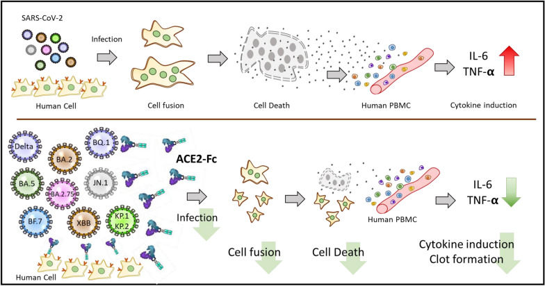
Background: The COVID-19 pandemic continues to affect the world in 2025. The rapid mutation of SARS-CoV-2 results in breakthrough infections and diminishes the efficacy of vaccines and anti-viral drugs. The severity of the disease varies across different variants, and the underlying mechanisms driving these differences remain unclear. This study explores the relationship between different Spike variants and cytotoxicity, aiming to determine whether the humanized decoy receptor ACE2-Fc can neutralize spikes from diverse variants, offering a solution to overcome rapid mutating SARS-CoV-2 induced immune escape.

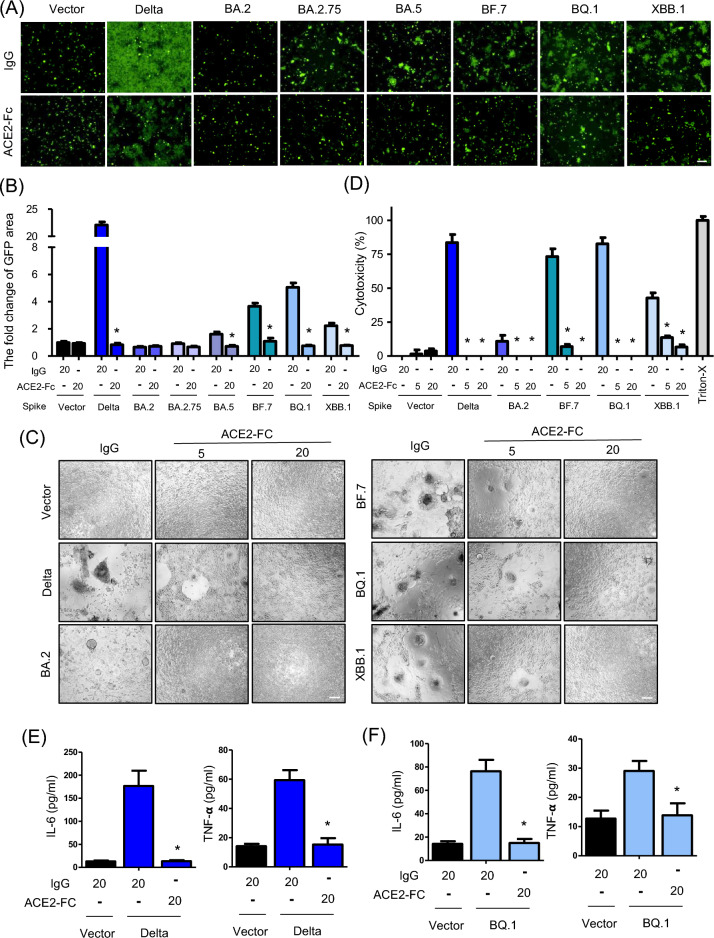
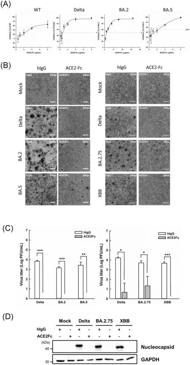
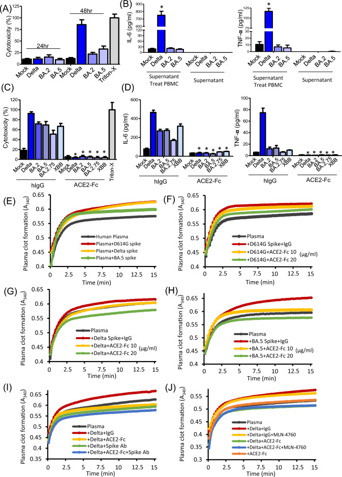
Methods: We co-cultured 293 T-ACE2 cells with 293 T cells transfected with various Spike protein variants or used H1650-ACE2 cells transfected with these Spike variants. This allowed us to observe the effects of different Spike mutations, specifically focusing on cell fusion, cytotoxicity, and cytokine release from human peripheral blood mononuclear cells. Flow cytometry is employed to determine if ACE2-Fc can recognize different Spike variants. We also assess the ability of ACE2-Fc to inhibit infection, cell fusion, cytotoxicity, and cytokine release through pseudovirus infections or Spike protein transfections. Additionally, we use actual viruses from SARS-CoV-2 patients to validate the impacts of Spike mutations and the effectiveness of ACE2-Fc. Furthermore, human plasma is utilized to evaluate ACE2-Fc's capability to inhibit Spike-induced clot formation.

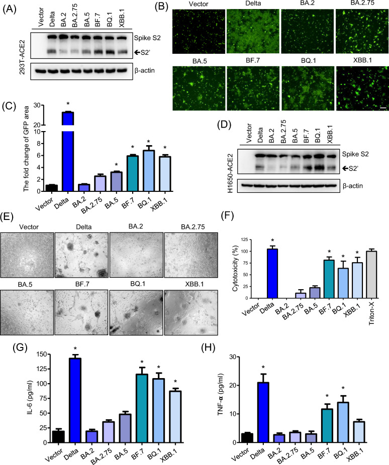
Results: We found that different Spike variants, particularly those with enhancements at the S2' site, increased cell-cell fusion capability, which correlated positively with cytotoxicity and cytokine IL-6 and TNF-α released from PBMCs. ACE2-Fc recognized spikes from wide-range of variants, including wild type, Alpha, Delta, Delta plus, Lambda, BA.2, BA.2.75, BA.5, BF.7, BQ.1, XBB.1, JN.1, KP.2, and KP.3, and effectively prevented these spike-expressing pseudo-viruses from entering host cells. Crucially, ACE2-Fc can prevent spike-induced cell fusion, thereby reducing subsequent cytotoxicity and the release of IL-6 and TNF-α from PBMCs. ACE2-Fc also effectively reduces plasma clot formation induced by trimeric spike proteins.

Conclusions: These findings demonstrated that ACE2-Fc could effectively combat the infection of rapidly mutating SARS-CoV-2, providing a potential solution to overcome immune evasion.

Keywords: ACE2-Fc; Immune escape; SARS-CoV-2; Spike.


Conflict of interest statement

Declarations. Ethics approval and consent to participate: Not applicable. Consent for publication: Not applicable. Competing interests: The authors declare no competing interests.

Figures

Fig. 1
Fig. 1
Varied properties of Spike protein variants. A The expression of different Spike variants in 293 T-ACE2 Cells. Two days after transfection, the presence of various Spike protein variants in cell lysates was confirmed by immunoblotting. β-actin was used as a loading control. B Cell Fusion in 293 T-ACE2 Cells. Images showing how different Spike protein variants induced cell fusion and syncytia formation. Scale bars represent 150 µm. C Quantitative syncytia analysis. The measured areas of syncytia formation to quantify the extent of cell fusion. D Levels of different Spike protein variants expressed in another cell line, H1650-ACE2. E Syncytia formation in H1650-ACE2 Cells. Similar to panel B, but demonstrating syncytia formation in H1650-ACE2 cells. Scale bars represent 150 µm. F Cytotoxicity post-Spike transfection. The cytotoxic effects observed 48 h after transfecting different Spike variants into H1650-ACE2 cells, with Triton-X100 treated cells serving as the 100% cytotoxicity control. G, H Impact on PBMCs. After treating PBMCs for 24 h with supernatants from H1650-ACE2 cells transfected with various Spike variants for 48 h, the levels of inflammation markers IL-6 and TNF-α were measured. Data are presented as mean ± SD from three replicates. Statistical analysis was performed by One-Way ANOVA with multiple comparisons *P < 0.05
Fig. 2
Fig. 2
Blocking of pseudovirus entry by ACE2-Fc across different Spike variants. A Recognition by ACE2-Fc. Flow cytometry analysis showing the binding of ACE2-Fc, tagged with a fluorescent marker (FITC), to different Spike protein variants expressed on 293 T cells. Mouse IgG-FITC was used as an isotype control to validate the specificity of ACE2-Fc binding. B, C Inhibition of Pseudovirus Entry. These panels demonstrate the effectiveness of ACE2-Fc in blocking the entry of pseudoviruses into two types of cells: 293 T-ACE2 (B) and H1650-ACE2 (C). The results indicate that ACE2-Fc can prevent pseudovirus infection by interfering with the interaction between the Spike protein and the ACE2 receptor on the surface of target cells. Data are presented as mean ± SD from three replicates. Statistical analysis was performed by unpaired two-tail t-test. *P < 0.05
Fig. 3
Fig. 3
Inhibitory effects of ACE2-Fc on Spike-induced cell fusion and cytotoxicity. A Syncytia formation in 293 T-ACE2 Cells. ACE2-Fc inhibits the formation of syncytia induced by different Spike protein variants. The effectiveness of the inhibition is visually represented, with scale bars measuring 150 µm. B Quantitative analysis of GFP area. The quantitative results of the green fluorescent (GFP) area, which reflects the extent of syncytia formation. The calculations are based on a formula detailed in the Methods section of the study. C Syncytia reversal in H1650-ACE2 Cells. ACE2-Fc can reverse syncytia formation caused by different Spike variants in another cell type, H1650-ACE2. Scale bars represent 150 µm. D Reduction of cytotoxicity. ACE2-Fc reduces cytotoxicity observed 48 h after transfecting different Spike variants into H1650-ACE2 cells. EF Reduction of cytokine induction. ACE2-Fc effectively reduces Delta and BQ.1 Spike meditated the induction of IL-6 and TNF-α in human PBMCs. Data are presented as mean ± SD from three replicates. Statistical analysis was performed by One-Way ANOVA with multiple comparisons (B, D) or unpaired two-tail t-test (E, F). *P < 0.05
Fig. 4
Fig. 4
Inhibition of SARS-CoV-2 entry into host cells by ACE2-Fc. A Plaque assay inhibition. The ability of ACE2-Fc to inhibit infection by different SARS-CoV-2 variants using a plaque assay. The effectiveness of ACE2-Fc is compared to human IgG (hIgG), which serves as a baseline for inhibition. B Yield reduction assay. This assay was conducted to evaluate the inhibitory effects of ACE2-Fc on various coronavirus variants in H1650-ACE2 cells. The assay measures the reduction in the number of infectious virus particles as a result of ACE2-Fc treatment. C Plaque assay in Vero-E6 cells. This plaque assay quantifies the viral titer in the supernatant collected from the yield reduction assay. This method assesses the amount of virus that remains infectious after treatment with ACE2-Fc. Data are presented as mean ± SD from three replicates. Statistical analysis was conducted using an unpaired two-tail t-test. *P < 0.05, **P < 0.01, ***P < 0.001. D Inhibition of nucleocapsid protein expression. The effect of ACE2-Fc on the expression of the Nucleocapsid protein across different variants. The results show a reduction in Nucleocapsid protein levels, indicating effective inhibition of virus replication by ACE2-Fc
Fig. 5
Fig. 5
Blocking of SARS-CoV-2 induced cytotoxicity, cytokine release and clot formation by ACE2-Fc. A The cytotoxicity in H1650-ACE2 cells. The cytotoxic effects observed in cells infected with different SARS-CoV-2 variants over 24 and 48 h. After the infection period, the supernatant was collected to measure cell damage, using Triton-X100 treated cells serving as the 100% cytotoxicity control. B Cytokine levels in PBMCs post-infection. After 48 h of infection in H1650-ACE2 cells, the supernatants were used to treat PBMCs, and the levels of IL-6 and TNF-α were measured. *Significant differences compared to the MOCK group. P < 0.05. C Inhibition of cytotoxicity by ACE2-Fc. Pre-treatment with ACE2-Fc significantly reduces the cytotoxic effects in H1650-ACE2 cells infected with various coronavirus variants 48 h post-infection. D Reduction of cytokine release by ACE2-Fc. ACE2-Fc treatment effectively decreases the release of IL-6 and TNF-α by PBMCs that were exposed to supernatants from infected H1650-ACE2 cells. Data are presented as mean ± SD from three replicates. Statistical analysis was performed by One-Way ANOVA with multiple comparisons *P < 0.05. E The turbidity of human plasma clot formation with D614G, Delta, and BA.5 spike. FH The effect of ACE2-Fc treatment on plasma clot formation induced by the D614G (F), Delta (G), and BA.5 H Spike proteins. I Effect of ACE2-Fc co-treatment with Spike antibody on plasma clot formation. (J) Effect of ACE2-Fc co-treatment with the ACE2 catalytic inhibitor MLN-4760 on plasma clot formation. EJ show representative results, with similar trends observed in three independent experiments
Fig. 6
Fig. 6
Schematic diagram of the impact of SARS-CoV-2 variants and ACE2-Fc inhibition. This diagram illustrates the process of cell fusion induced by different SARS-CoV-2 variants, which leads to cytotoxicity and cytokine induction in host cells. It also shows how ACE2-Fc treatment can inhibit these effects. ACE2-Fc is highlighted as a therapeutic agent that effectively blocks the disease progression at multiple stages: preventing the virus from entering cells, reducing cell fusion, mitigating cell damage, and decreasing the release of inflammatory cytokines

Similar articles

References

    1. Wu F, Zhao S, Yu B, Chen YM, Wang W, Song ZG, Hu Y, Tao ZW, Tian JH, Pei YY, et al. A new coronavirus associated with human respiratory disease in China. Nature. 2020;579(7798):265–9. - PMC - PubMed
    1. Liu WJ, Liu P, Lei W, Jia Z, He X, Shi W, Tan Y, Zou S, Wong G, Wang J, et al. Surveillance of SARS-CoV-2 at the huanan seafood market. Nature. 2024;631(8020):402–8. - PubMed
    1. Steiner S, Kratzel A, Barut GT, Lang RM, Aguiar Moreira E, Thomann L, Kelly JN, Thiel V. SARS-CoV-2 biology and host interactions. Nat Rev Microbiol. 2024;22(4):206–25. - PubMed
    1. Huang KY, Lin MS, Yang PC. Using host receptor as a decoy to treat COVID-19: a solution for immune escape? EMBO Mol Med. 2022;14(11): e16818. - PMC - PubMed
    1. Lugano D, Kutima B, Kimani M, Sigilai A, Gitonga J, Karani A, Akech D, Karia B, Ziraba AK, Maina A, et al. Evaluation of population immunity against SARS-CoV-2 variants, EG.5.1, FY.4, BA.2.86, JN.1, JN.1.4, and KP.3.1.1 using samples from two health demographic surveillance systems in Kenya. BMC Infect Dis. 2024;24(1):1474. - PMC - PubMed

MeSH terms

Supplementary concepts