

Antitrypanosomal potential of Salvia officinalis terpenoids-rich fraction in Trypanosoma evansi-infected rat model
- PMID: 40571943
- PMCID: PMC12203732
- DOI: 10.1186/s12917-025-04861-2
Antitrypanosomal potential of Salvia officinalis terpenoids-rich fraction in Trypanosoma evansi-infected rat model
Abstract
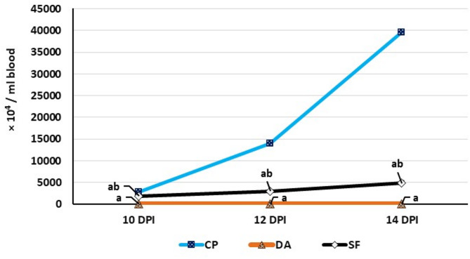
BACKGROUND TRYPANOSOMA EVANSI : (T. evansi) is a major protozoan disease that affects animals, including camels, and causes substantial economic detriments. The failure to control T. evansi infections is due to the unavailability of vaccines and the development of resistance to existing chemical drugs. In this study, we evaluated the effect of Salvia officinalis terpenoids-rich fraction on the degree of parasitemia and associated pathological alterations in rats experimentally infected with T. evansi.
Method: Eighty adult male rats were equally divided into 4 groups. The first group was a negative control. The second group was intraperitoneally infected with T. evansi at a dose of 1 × 10⁴ trypanosomes. The third group was similarly infected and subsequently treated intramuscularly with diminazene aceturate at a dose of 7 mg/kg body weight (b.wt.). The fourth group received a daily oral administration of Salvia officinalis terpenoids-rich fraction at a dose of 300 mg/kg b.wt. throughout the experimental period and was also infected with T. evansi.
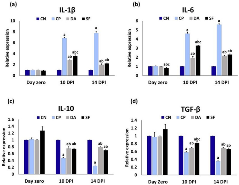
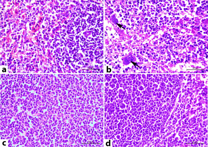
Result: The infection with T. evansi resulted in normocytic normochromic anemia, leukocytosis, hypoglycemia, hypertriglyceridemia, an increase in very low-density lipoprotein (VLDL) cholesterol, and reductions in high-density lipoprotein (HDL) and low-density lipoprotein (LDL) cholesterols. Additionally, the infection induced upregulation of the pro-inflammatory cytokines such as interleukin-1beta (IL-1β) and interleukin-6 (IL-6), and downregulation of the anti-inflammatory cytokines such as interleukin-10 (IL-10) and transforming growth factor beta (TGF-β). Besides histopathological changes in the brain and spleen, T. evansi markedly elevated brain oxidative stress and acetylcholinesterase (AChE) activity. The treatment with salvia fraction significantly decreased the degree of parasitemia and mitigated the T. evansi-induced pathological alterations.
Conclusion: The terpenoids-rich fraction from Salvia officinalis exhibits antitrypanosomal activity and may serve as a promising candidate for developing novel trypanocidal agents.
Keywords: Salvia officinalis; Trypanosome evansi; Antitrypanosomal activity; Terpenoids.
© 2025. The Author(s).
Conflict of interest statement
Declarations. Ethical approval: The whole experiment was conducted according to Animal Research Ethics Guidelines at the Faculty of Veterinary Medicine, Beni-Suef University, Egypt, with approval number 022–296. Consent to participate: Not applicable. Consent for publication: Not applicable. Competing interests: The authors declare no competing interests.
Figures


Similar articles
-
Clinical occurrence of trypanosomiasis in Arabian horses from Ahvaz.Vet Clin Pathol. 2025 Jun;54(2):186-190. doi: 10.1111/vcp.70003. Epub 2025 Jun 3. Vet Clin Pathol. 2025. PMID: 40462454
-
Corticosteroids for the treatment of Duchenne muscular dystrophy.Cochrane Database Syst Rev. 2016 May 5;2016(5):CD003725. doi: 10.1002/14651858.CD003725.pub4. Cochrane Database Syst Rev. 2016. PMID: 27149418 Free PMC article.
-
Systemic pharmacological treatments for chronic plaque psoriasis: a network meta-analysis.Cochrane Database Syst Rev. 2017 Dec 22;12(12):CD011535. doi: 10.1002/14651858.CD011535.pub2. Cochrane Database Syst Rev. 2017. Update in: Cochrane Database Syst Rev. 2020 Jan 9;1:CD011535. doi: 10.1002/14651858.CD011535.pub3. PMID: 29271481 Free PMC article. Updated.
-
A Survey on Trypanosoma evansi (Kinetoplastida, Trypanosomatidae) Infection in Domestic Animals in a Surra Endemic Area of Southern Algeria.Vector Borne Zoonotic Dis. 2024 Apr;24(4):219-225. doi: 10.1089/vbz.2023.0015. Epub 2024 Feb 27. Vector Borne Zoonotic Dis. 2024. PMID: 38416509
-
Sertindole for schizophrenia.Cochrane Database Syst Rev. 2005 Jul 20;2005(3):CD001715. doi: 10.1002/14651858.CD001715.pub2. Cochrane Database Syst Rev. 2005. PMID: 16034864 Free PMC article.
References
-
- Khalafalla AI, Hussein MF, Bornstein S. Trypanosomosis. Infectious Diseases of Dromedary Camels: A Concise Guide 2021:273–296.10.1007/978-3-030-79389-0_46
-
- Ata EB, Abdel-Aziz TH, Abdel-Ghany HS, Elsawy BS, Abdullah HH, Abouelsoued D, et al. Molecular and serological diagnosis of the circulating Trypanosoma evansi in Egyptian livestock with risk factors assessment. Microb Pathog. 2024;197107073. 10.1016/j.micpath.2024.107073 - PubMed
-
- Kim J, Álvarez-Rodríguez A, Li Z, Radwanska M, Magez S. Recent progress in the detection of surra, a neglected disease caused by Trypanosoma evansi with a one health impact in large parts of the tropic and sub-tropic world. Microorganisms. 2023;12(1):44. 10.3390/microorganisms12010044 - PMC - PubMed
MeSH terms
Substances
LinkOut - more resources
Full Text Sources
