. 2025 May 23;13(6):1189.
doi: 10.3390/microorganisms13061189.

Chlorophyllin-Mediated Photodynamic Inactivation: Dosage and Time Dependency in the Inhibition of Bacillus subtilis

Affiliations

Chlorophyllin-Mediated Photodynamic Inactivation: Dosage and Time Dependency in the Inhibition of Bacillus subtilis

Sarah Pohland et al. Microorganisms. .

Abstract

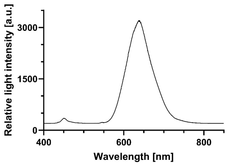
Photodynamic inactivation of bacteria offers a promising alternative to counteract the trend towards the development of resistance, which, if left uncontrolled, will lead to the death of 10 million people per year by 2050. Its advantage over antibiotics is the site-specific mode of action due to the photosensitizer (PS) and the low risk of developing resistance. This is primarily prevented by the damage of the bacteria, which also destroy internal structures such as nucleic acid, proteins, and lipids. A promising and still little-researched PS is chlorophyllin (CHL), a chlorophyll derivative. This study investigated its mode of action on Bacillus subtilis growth using optical density (OD) measurements. It was shown that the PS is highly effective even at low concentrations and short irradiation durations. Here, 1 mg/L and an irradiation duration of 1 min were sufficient to inhibit the growth of the Gram-positive bacterium Bacillus subtilis for several hours.

Keywords: antibiotic alternatives; chlorophyllin; photodynamic inactivation (PDI); photodynamic therapy (PDT); photosensitizer.


Conflict of interest statement

Author Michael Lebert is engaged with the company Space Biology Unlimited, SAS. There is no conflict of interest regarding this MS. The remaining authors declare that the research was conducted in the absence of any commercial or financial relationships that could be construed as a potential conflict of interest.

Figures

Figure 1
Figure 1
Emission spectrum of red light source for irradiation of B. subtilis.
Figure 2
Figure 2
Effect of chlorophyllin and influence of light exposure on bacterial growth. B. subtilis was incubated during the exponential phase with 1 mg/L CHL or in LB medium (control). Light exposure for one minute (left) and absence of light (right). Optical density was measured every 20 min for a period of 20 h. The data consist of three replicates (n = 3) of the experiment, using the average from each replicate of three technical replicates, including their standard deviation.
Figure 3
Figure 3
Volcano plots for the measurement data from Figure 2. One-minute exposure resulted in a statistically significant reduction in growth rate after CHL treatment at each measurement time (left), while lack of exposure despite CHL treatment had no statistically significant effect on growth (right). Each data point represents the mean difference and corresponding −log10(p-value) from unpaired t-tests. The horizontal line indicates the significance threshold at p = 0.05. No correction for multiple comparisons was applied.
Figure 4
Figure 4
Effect of treatment of B. subtilis with chlorophyllin (CHL) 1 mg/L and red light 899 µmol photons·m2·s−1 for 1 min on the survival rate of B. subtilis. Control: light control without CHL; CHL: cells incubated with chlorophyllin during illumination. ***: p-value < 0.001.
Figure 5
Figure 5
Effect of chlorophyllin (1 mg/L) and (A) light (899 µmol photons·m2·s−1 for 1 min) or in (B) darkness, respectively, on germination and growth of B. subtilis spores. Statistical comparison between groups was performed using unpaired t-tests at individual time points (C,D). Significance was defined as p < 0.05. Data are shown as mean ± SD.
Figure 6
Figure 6
CFUs after plating CHL-incubated cells after light exposure (899 µmol photons·m2·s−1 for 1 min, DPI 0.14 mol/m2) or darkness, respectively. Control: Cells in absence of light and no CHL treatment. CHL+/L+: Cells incubated in CHL in presence of light. CHL+/L: Cells with CHL incubated in darkness (ns: not significant).
Figure 7
Figure 7
Effect of chlorophyllin and influence of light exposure on bacterial growth. B. subtilis with spores was incubated with 10 mg/L CHL or in LB medium (control). (A) Light exposure for one minute; (B) darkened for fifteen minutes. Optical density was measured every 30 min over several hours. Data consist of two replicates (n = 2) of experiment, using average from each replicate of two technical replicates, including their standard deviation. (C) One-minute exposure resulted in statistically significant reduction in growth rate after CHL treatment at most measurement times; (D) data set of chlorophyllin-treated bacteria under dark conditions shows no significant differences; statistical comparison between groups was performed using unpaired t-tests at individual time points. Significance was defined as p < 0.05. Data are shown as mean ± SD.
Figure 8
Figure 8
Effect of chlorophyllin (20 mg/L) and light exposure on growth of spore-containing B. subtilis; (A) growth after 15 min light exposure; (B) growth after 15 min in darkness; (C) volcano plot of data from light-exposed bacteria: light-activated Chlorophyllin exerts significant effect on growth; (D) statistical comparison between groups was performed using unpaired t-tests at individual time points. Significance was defined as p < 0.05. Data are shown as mean ± SD.
Figure 9
Figure 9
Effect of chlorophyllin (CHL) and light (15.3 µmol photons·m2·s−1) on transcription level of ROS-related genes recA, uvrA, and mfd, respectively. Data show relative expression compared to gyrA as reference gene. Left column: light-treated samples; right column: dark exposed samples. +control: positive control with 150 µM H2O2; −control: negative control: untreated cells; CHL: cells incubated in presence of chlorophyllin. N = 3, with each 3 technical triplicates. Statistical analysis performed with unifactorial ANOVA with Holm–Šídák correction (ns: not significant, *: p < 0.033, ** p < 0.002, *** p < 0.001).
Figure 10
Figure 10
Increase in glutathione peroxidase (GPX) activity in light and darkness induced by chlorophyllin (CHL). Light alone did not alter GPX-activity. Negative controls were without CHL; light exposed samples (L+) were illuminated with 15.3 µmol photons·m2·s−1 of red light for 5 s. N = 4, with each 3 technical replicates. Statistical analysis performed with unifactorial ANOVA with Holm–Šídák correction. (****: p-value < 0.001).
Figure 11
Figure 11
Effect of chlorophyllin (CHL) and light on catalase-activity. B. subtilis-cells were incubated in 1 mg/L CHL and exposed to red light (5 s, 15.3 µmol photons·m2·s−1)-CHL+/L+. Controls were dark-exposed cells with CHL (CHL+/L), illuminated cells without CHL (CHL/L+), dark-exposed cells with CHL (CHL+/L), and a kit-internal positive control +control. N = 3 with 3 technical triplicates. Statistical analysis performed with unifactorial ANOVA with Holm–Šídák correction. (ns: not significant, *: p < 0.033, *** p < 0.001).
Figure 12
Figure 12
Superoxide dismutase activity is not significantly affected by chlorophyllin, 1 mg/L red light illumination (15.3 µmol photons·m2·s−1, 5 s), or combination of these factors. CHL/L+ (no chlorophyllin/light); CHL+/L+ (with chlorophyllin/light); CHL/L (no chlorophyllin/dark); CHL+/L (chlorophyllin/dark) (ns: not significant).

Similar articles

References

    1. Larsson D.G.J., Flach C.-F. Antibiotic resistance in the environment. Nat. Rev. Microbiol. 2022;20:257–269. doi: 10.1038/s41579-021-00649-x. - DOI - PMC - PubMed
    1. Pulingam T., Parumasivam T., Gazzali A.M., Sulaiman A.M., Chee J.Y., Lakshmanan M., Chin C.F., Sudesh K. Antimicrobial resistance: Prevalence, economic burden, mechanisms of resistance and strategies to overcome. Eur. J. Pharm. Sci. 2022;170:106103. doi: 10.1016/j.ejps.2021.106103. - DOI - PubMed
    1. O’Neill J. Review on Antimicrobial Resistance. Antimicrobial Resistance: Tackling a Crisis for the Health and Wealth of Nations. Volume 2014. Review on Antimicrobial Resistance; London, UK: 2014. pp. 1–16.
    1. Kolarikova M., Hosikova B., Dilenko H., Barton-Tomankova K., Valkova L., Bajgar R., Malina L., Kolarova H. Photodynamic therapy: Innovative approaches for antibacterial and anticancer treatments. Med. Res. Rev. 2023;43:717–774. doi: 10.1002/med.21935. - DOI - PubMed
    1. Donohoe C., Senge M.O., Arnaut L.G., Gomes-da-Silva L.C. Cell death in photodynamic therapy: From oxidative stress to anti-tumor immunity. Biochim. Biophys. Acta Rev. Cancer. 2019;1872:188308. doi: 10.1016/j.bbcan.2019.07.003. - DOI - PubMed

LinkOut - more resources