

Epicardial Adipose Tissue: A Multimodal Imaging Diagnostic Perspective
- PMID: 40572649
- PMCID: PMC12195245
- DOI: 10.3390/medicina61060961
Epicardial Adipose Tissue: A Multimodal Imaging Diagnostic Perspective
Abstract
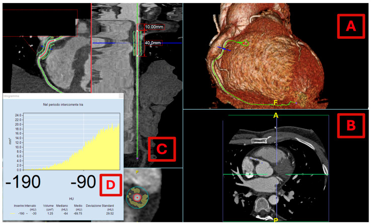
Epicardial adipose tissue (EAT), strategically located between the myocardium and the visceral pericardial layer, is increasingly recognized as an active player in cardiovascular health rather than a passive fat depot. EAT secretes a notable array of bioactive molecules known as adipokines, which exert critical exocrine and paracrine effects. Recent research has focused on pericoronary adipose tissue (PCAT)-the EAT surrounding coronary arteries-demonstrating its intricate bidirectional relationship with the vascular wall. Under normal physiological conditions, this interaction promotes vascular homeostasis; however, dysfunctional PCAT can release pro-inflammatory adipokines implicated in the pathogenesis of atherogenesis. Notably, PCAT inflammation has emerged as a significant factor associated with the development of coronary artery disease (CAD) and major cardiovascular events. This review seeks to elucidate the imaging methodologies employed to evaluate EAT, emphasizing cardiac computed tomography (CCT) as the preeminent imaging modality. Unlike echocardiography and cardiac magnetic resonance imaging, CCT not only visualizes and quantifies EAT but also concurrently assesses coronary arteries and PCAT. Recent findings have established the potential of CCT-derived PCAT attenuation as a noninvasive biomarker for coronary inflammation, offering prospects for monitoring therapeutic responses to innovative anti-inflammatory interventions in CAD management.
Keywords: echocardiography; epicardial adipose tissue; multimodality imaging; pericoronary adipose tissue.
Conflict of interest statement
The authors declare no conflicts of interest.
Figures


References
-
- Romano A.D., La Marca A., Villani R., Sangineto M., Manuppelli V., Brunetti N.D., Vendemiale G., Serviddio G. Exploring the Relationship between Epicardial Fat Thickness and Coronary Revascularization: Implications for Cardiovascular Health. J. Clin. Med. 2023;13:247. doi: 10.3390/jcm13010247. - DOI - PMC - PubMed
-
- Guglielmo M., Penso M., Carerj M.L., Giacari C.M., Volpe A., Fusini L., Baggiano A., Mushtaq S., Annoni A., Cannata F., et al. DEep LearnIng-Based QuaNtification of Epicardial Adipose Tissue Predicts MACE in Patients Undergoing Stress CMR. Atherosclerosis. 2024;397:117549. doi: 10.1016/j.atherosclerosis.2024.117549. - DOI - PubMed
Publication types
MeSH terms
LinkOut - more resources
Full Text Sources
Miscellaneous
