

Comparative Analysis of Corneal Morphological and Optical Parameters in Predicting DSAEK Surgery Outcome
- PMID: 40572712
- PMCID: PMC12195242
- DOI: 10.3390/medicina61061022
Comparative Analysis of Corneal Morphological and Optical Parameters in Predicting DSAEK Surgery Outcome
Abstract
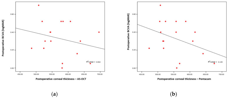
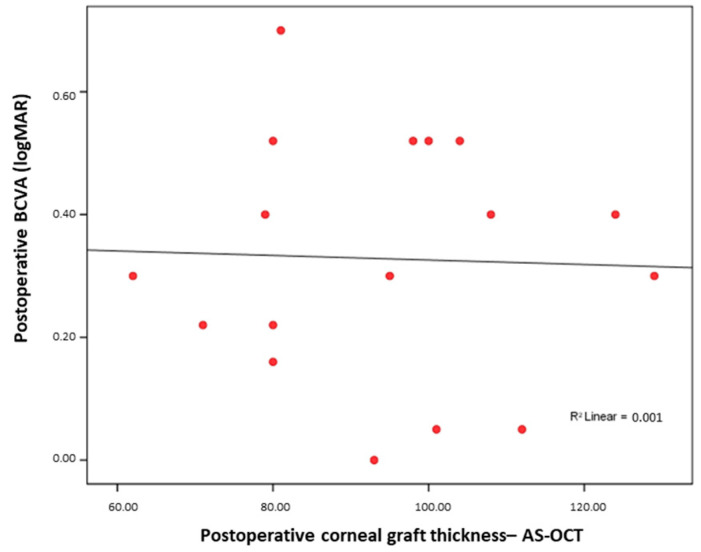
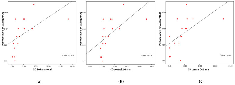
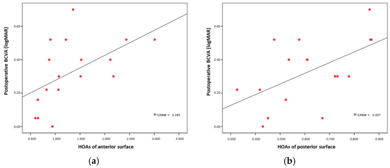
Background and Objectives: Descemet stripping automated endothelial keratoplasty (DSAEK) is a widely used surgical technique for treating corneal endothelial dysfunctions such as Fuchs endothelial corneal dystrophy (FECD) and pseudophakic bullous keratopathy (PBK). This study aimed to investigate the association between postoperative visual acuity and various corneal morphological and optical parameters, including corneal densitometry (CD) and higher-order aberrations (HOAs), measured using the Pentacam (OCULUS Optikgeräte GmbH, Wetzlar, Germany), as well as graft thickness, which was assessed by anterior segment optic coherence tomography (AS-OCT), (Optovue Inc., Fremont, CA, USA), and corneal thickness, assessed by both AS-OCT and Pentacam. Materials and Methods: This prospective, observational cohort study included 17 eyes from 13 patients who underwent DSAEK. Data on preoperative visual acuity were collected, while postoperative parameters were assessed during follow-up visits. Corneal measurements included the total corneal and corneal graft thickness, corneal densitometry in 20 defined subregions, and corneal higher-order aberrations. Associations between these parameters and postoperative visual acuity were evaluated using nonparametric statistical tests. Results: The postoperative visual acuity improved significantly (p < 0.001). Strong correlations were found between poorer visual acuity and higher CD values. The strongest correlations with visual acuity were found for CD 2-6 mm total (Rho = 0.795; p < 0.001), CD central 2-6 mm (Rho = 0.791; p < 0.001), and CD central 0-2 mm (Rho = 0.730; p < 0.001). Significant associations were also observed with anterior and posterior HOAs (Rho = 0.624, p = 0.01; and Rho = 0.556, p = 0.02, respectively). No correlation was found between visual outcomes and graft thickness measured by AS-OCT (Rho = 0.051; p = 0.85), nor with total corneal thickness measured by AS-OCT (Rho = -0.227; p = 0.38) or Pentacam (Rho = -0.369; p = 0.14). Conclusions: This study demonstrates that CD and HOAs are more strongly associated with postoperative visual acuity after DSAEK than traditionally monitored parameters such as graft or corneal thickness. The results highlight the value of detailed corneal imaging and support the use of advanced optical diagnostics in postoperative evaluation.
Keywords: Descemet stripping automated endothelial keratoplasty; cornea; corneal imaging; corneal transplantation.
Conflict of interest statement
The authors declare no conflicts of interest.
Figures
Similar articles
-
Mediators of Visual Acuity in Descemet Membrane Endothelial Keratoplasty and Ultrathin Descemet Stripping Automated Endothelial Keratoplasty.Cornea. 2024 Jul 1;43(7):853-859. doi: 10.1097/ICO.0000000000003434. Epub 2023 Nov 21. Cornea. 2024. PMID: 37986182 Clinical Trial.
-
Descemet's membrane endothelial keratoplasty (DMEK) versus Descemet's stripping automated endothelial keratoplasty (DSAEK) for corneal endothelial failure.Cochrane Database Syst Rev. 2018 Jun 25;6(6):CD012097. doi: 10.1002/14651858.CD012097.pub2. Cochrane Database Syst Rev. 2018. PMID: 29940078 Free PMC article.
-
Analysis of Corneal Scheimpflug Densitometry and Ocular Wavefront Aberrations Post Descemet Stripping Automated Endothelial Keratoplasty.Eye Contact Lens. 2022 Jun 1;48(6):242-249. doi: 10.1097/ICL.0000000000000868. Epub 2021 Nov 25. Eye Contact Lens. 2022. PMID: 34860724
-
Endothelial keratoplasty versus penetrating keratoplasty for Fuchs endothelial dystrophy.Cochrane Database Syst Rev. 2011 Jul 6;(7):CD008420. doi: 10.1002/14651858.CD008420.pub2. Cochrane Database Syst Rev. 2011. Update in: Cochrane Database Syst Rev. 2014 Feb 14;(2):CD008420. doi: 10.1002/14651858.CD008420.pub3. PMID: 21735430 Updated.
-
Modified Method for Nanothin Descemet Stripping Automated Endothelial Keratoplasty.Cornea. 2024 Dec 1;43(12):1522-1527. doi: 10.1097/ICO.0000000000003507. Cornea. 2024. PMID: 38381043
References
Publication types
MeSH terms
LinkOut - more resources
Full Text Sources
Research Materials
