. 2025 Jun 7;18(12):2690.
doi: 10.3390/ma18122690.

Differential lncRNA Expression in Undifferentiated and Differentiated LUHMES Cells Following Co-Exposure to Silver Nanoparticles and Nanoplastic

Affiliations

Differential lncRNA Expression in Undifferentiated and Differentiated LUHMES Cells Following Co-Exposure to Silver Nanoparticles and Nanoplastic

Kamil Brzóska et al. Materials (Basel). .

Abstract

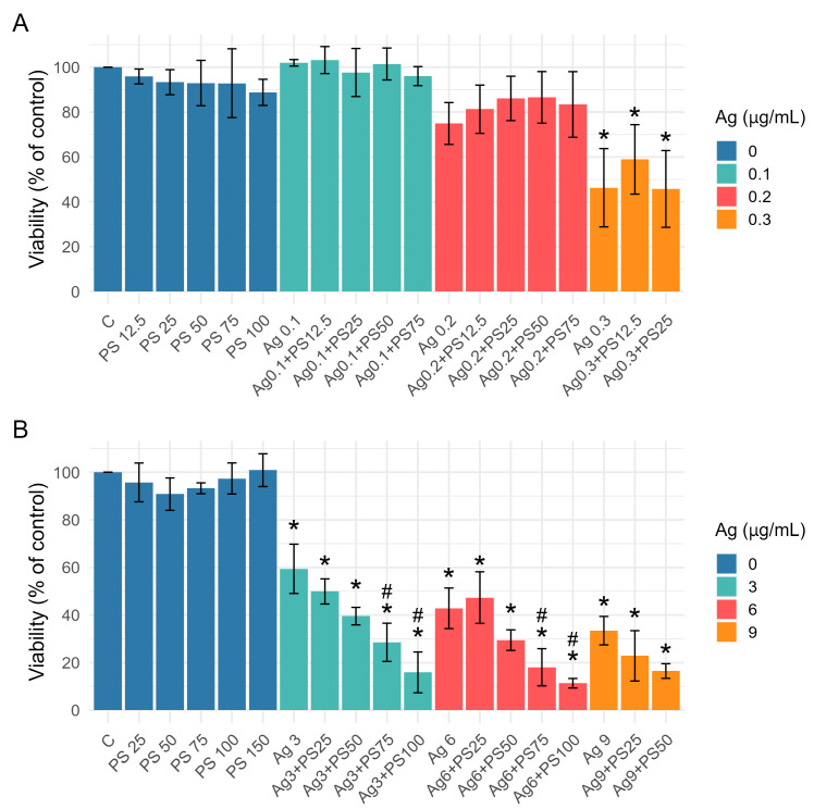
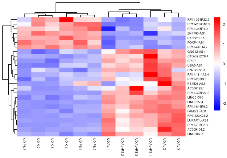
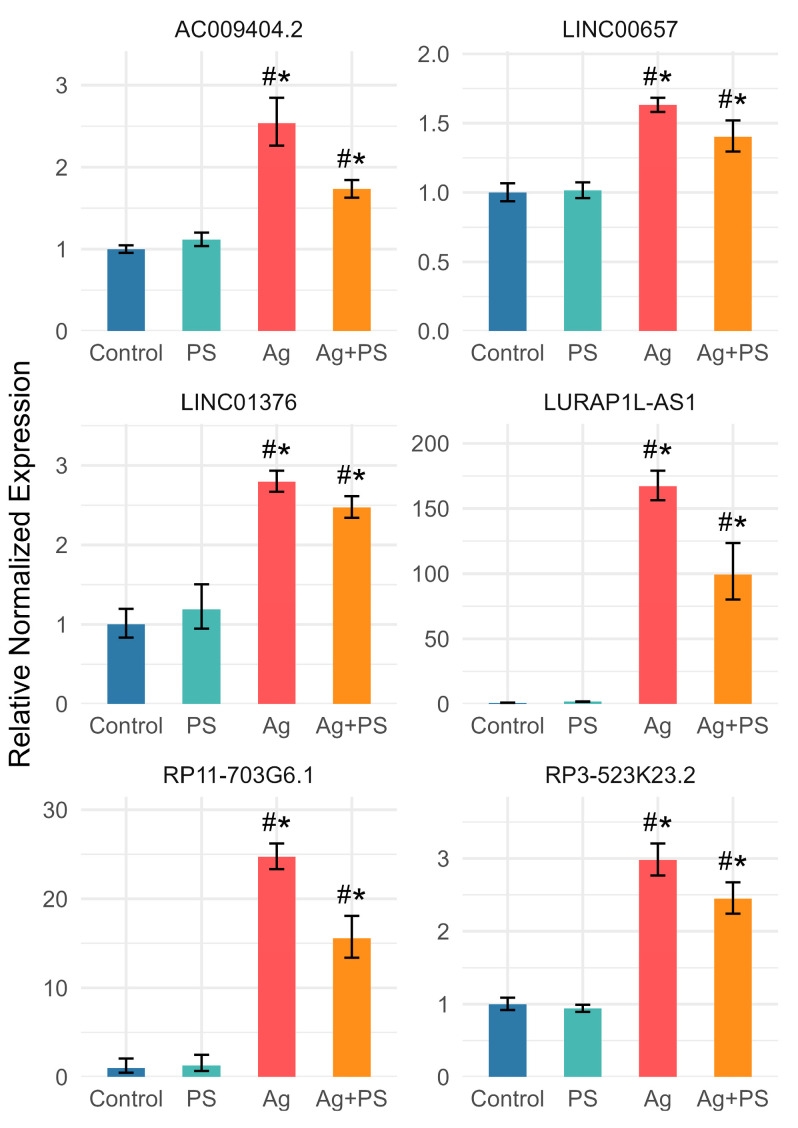
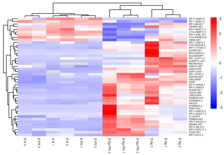
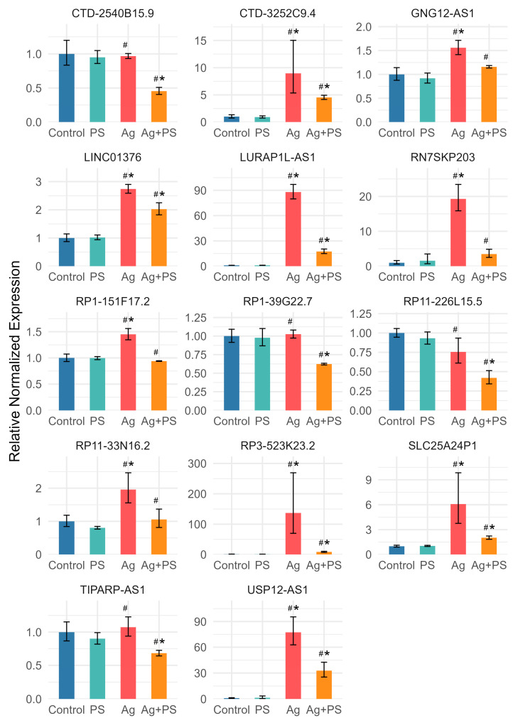
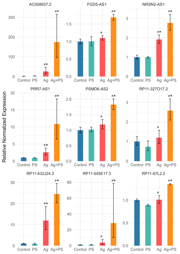
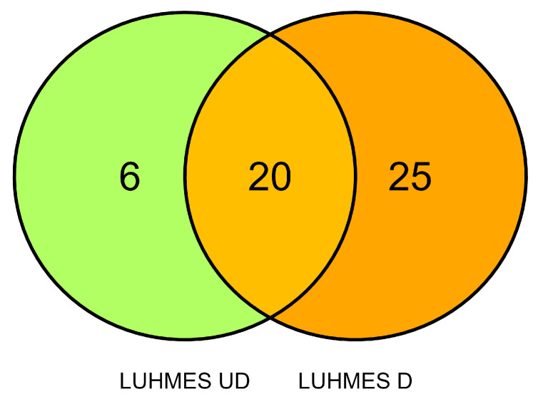
Human exposure to micro- and nanoplastic (MNP) has become an increasing concern due to its accumulation in the environment and human body. In the human organism, MNP accumulates in various tissues, including the central nervous system, where it is associated which neurotoxic effects. Beyond its inherent toxicity, MNP also acts as a carrier for various chemical contaminants, including metals. Consequently, recent studies emphasize the importance of the evaluation of co-exposure scenarios involving MNP and other types of nanoparticles. In this study, we investigated effects of co-exposure to 20 nm silver nanoparticles (AgNPs) and 20 nm polystyrene nanoparticles (PSNPs) on cell viability and the expression of inflammation-related long non-coding RNAs (lncRNAs) in undifferentiated and differentiated Lund human mesencephalic (LUHMES) cells. While PSNPs alone did not significantly affect cell viability or lncRNA expression, AgNPs markedly reduced viability and deregulated lncRNA expression in both cell types. Notably, in differentiated cells, co-exposure to AgNPs and high concentrations of PSNPs led to a significantly greater reduction in viability compared to AgNPs alone, suggesting a synergistic effect. At the molecular level, both synergistic and antagonistic interactions between AgNPs and PSNPs were observed in the regulation of lncRNA expression, depending on the cell differentiation status. These findings highlight the complex biological interactions between AgNPs and PSNPs and emphasize the importance of considering nanoparticle co-exposures in toxicological evaluations, as combined exposures may significantly affect cellular and molecular responses.

Keywords: AgNPs; LUHMES cells; lncRNA; nanoplastic; silver nanoparticles.


Conflict of interest statement

The authors declare no conflicts of interest. The funders had no role in the design of the study; in the collection, analyses, or interpretation of data; in the writing of the manuscript; or in the decision to publish the results.

Figures

Figure 1
Figure 1
HR-SEM images of AgNPs (A,B) and PSNPs (C,D) used in the study.
Figure 2
Figure 2
Viability of undifferentiated (A) and differentiated (B) LUHMES cells following exposure to AgNPs, PSNPs, or their combination for 24 h. Bars represent mean values, with error bars indicating the standard deviation from three independent experiments. Asterisks (*) denote statistically significant differences compared to control group (post-hoc Tukey test), while hash symbols (#) indicate statistically significant differences compared to the AgNP-treated group (post-hoc Tukey test).
Figure 3
Figure 3
Heatmap of lncRNAs significantly deregulated in undifferentiated LUHMES cells after exposure to AgNPs or their mixture with PSNPs. Expression values are standardized per gene (row Z-score).
Figure 4
Figure 4
Expression of lncRNAs for which PSNPs suppressed AgNP-induced upregulation in undifferentiated LUHMES cells. Bars represent mean values, with error bars indicating the standard deviation from three independent experiments. Asterisks (*) denote statistically significant differences compared to control group (post-hoc Tukey test), while hash symbols (#) indicate statistically significant differences compared to the AgNP-treated group (post-hoc Tukey test).
Figure 5
Figure 5
Heatmap of lncRNAs significantly deregulated in differentiated LUHMES cells after exposure to AgNPs or their mixture with PSNPs. Expression values are standardized per gene (row Z-score).
Figure 6
Figure 6
Expression of lncRNAs for which PSNPs suppressed AgNP-induced upregulation in differentiated LUHMES cells. Bars represent mean values, with error bars indicating the standard deviation from three independent experiments. Asterisks (*) denote statistically significant differences compared to control group (post-hoc Tukey test), while hash symbols (#) indicate statistically significant differences compared to the AgNP-treated group (post-hoc Tukey test).
Figure 7
Figure 7
Expression of lncRNAs for which synergy of PSNPs and AgNPs was observed in differentiated LUHMES cells. Bars represent mean values, with error bars indicating the standard deviation from three independent experiments. Asterisks (*) denote statistically significant differences compared to control group (post-hoc Tukey test), while hash symbols (#) indicate statistically significant differences compared to the AgNP-treated group (post-hoc Tukey test).
Figure 8
Figure 8
Venn diagram comparing lncRNA deregulation in undifferentiated (UD) and differentiated (D) LUHMES cells.

Similar articles

References

    1. Zahra Z., Habib Z., Hyun S., Sajid M. Nanowaste: Another Future Waste, Its Sources, Release Mechanism, and Removal Strategies in the Environment. Sustainability. 2022;14:2041. doi: 10.3390/su14042041. - DOI
    1. Shukla S., Khanna S., Khanna K. Unveiling the Toxicity of Micro-Nanoplastics: A Systematic Exploration of Understanding Environmental and Health Implications. Toxicol. Rep. 2025;14:101844. doi: 10.1016/j.toxrep.2024.101844. - DOI - PMC - PubMed
    1. Toussaint B., Raffael B., Angers-Loustau A., Gilliland D., Kestens V., Petrillo M., Rio-Echevarria I.M., Van den Eede G. Review of Micro- and Nanoplastic Contamination in the Food Chain. Food Addit. Contam. Part. A. 2019;36:639–673. doi: 10.1080/19440049.2019.1583381. - DOI - PubMed
    1. Waring R.H., Harris R.M., Mitchell S.C. Plastic Contamination of the Food Chain: A Threat to Human Health? Maturitas. 2018;115:64–68. doi: 10.1016/j.maturitas.2018.06.010. - DOI - PubMed
    1. Li H., Zhu L., Ma M., Wu H., An L., Yang Z. Occurrence of Microplastics in Commercially Sold Bottled Water. Sci. Total Environ. 2023;867:161553. doi: 10.1016/j.scitotenv.2023.161553. - DOI - PubMed

LinkOut - more resources