Review
. 2025 Jun 12;18(12):2752.
doi: 10.3390/ma18122752.

Decellularized Extracellular Matrices for Skin Wound Treatment

Affiliations
Review

Decellularized Extracellular Matrices for Skin Wound Treatment

Rui Liang et al. Materials (Basel). .

Abstract

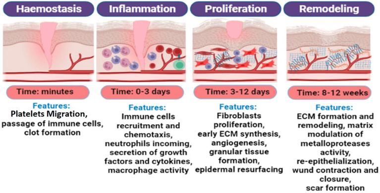
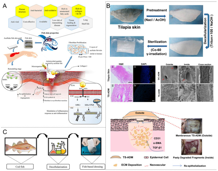
Skin trauma, especially chronic trauma, poses a significant clinical challenge, often leading to severe disability or even death. Traditional treatment methods exhibit several limitations in terms of efficacy, material availability, and biocompatibility. The development of decellularized extracellular matrices (dECMs) has led to revolutionary progress in this field. These materials retain the bioactive components of the natural extracellular matrix (ECM) and, combined with their excellent physical structure, promote wound healing. Preclinical studies have demonstrated that dECM-based dressings can enhance the re-epithelialization rate by 20-50% and shorten the healing cycle of chronic wounds by 40%. This article systematically reviews the application of dECM in wound repair. First, it outlines the pathophysiology of wound healing, focusing on the mechanisms by which key ECM components promote wound healing. Next, it classifies decellularized materials and proposes material design schemes for different types of damage. Finally, the limitations of current dECM-based wound treatments and future research directions are discussed. This review aims to provide a theoretical framework and technical reference for researchers in related fields, promoting the widespread application of dECM materials for skin trauma treatment.

Keywords: biomaterials; decellularized matrix; extracellular matrix; skin trauma; wound healing.


Conflict of interest statement

The authors declare no conflict of interest.

Figures

Figure 1
Figure 1
The different stages of the wound-healing process and their characteristics. Reprinted with permission from Ref. [40]. Copyright 2022 MDPI.
Figure 2
Figure 2
Acellular dermal matrix (ADM) biomaterials for wound healing. (A) The construction of a human skin bioscaffold (hSBS) after the decellularization of human natural skin tissue (hNST) and the proportion of classes constituting matrix bodies in hNSTs and hSBSs. Reprinted with permission from Ref. [130]. Copyright 2020 American Chemical Society. (B) The number and proportion of matrix body proteins in human and porcine skin bioscaffolds. Reprinted with permission from Ref. [130]. Copyright 2020 American Chemical Society. (C) A schematic of synthetic reduced graphene oxide (RGO) combined with ADMs composite scaffolds loaded with MSCs for use in a mouse diabetic wound-healing model, and scanning electron microscope (SEM) images of the three scaffolds. Reprinted with permission from Ref. [137]. Copyright 2019 American Chemical Society. (D) The mechanism of ADM-Fe3+@PA-Exos/GelMA promoting diabetic wound repair and images of antimicrobial experiments. Reprinted with permission from Ref. [138]. Copyright 2023 Elsevier. Abbreviation: ADM-Fe3+@PA-Exos/GelMA: ADM modified by trivalent iron ion (Fe3+) @ protocatechualdehyde (PA) complex, gelatin methacrylate (GelMA), and exosomes.
Figure 4
Figure 4
Decellularized small intestinal submucosa (DSIS) for wound healing. (A) Decellularization process of porcine SIS. Reprinted with permission from Ref. [159]. Copyright 2023 Elsevier. (B) H&E tissue sections of decellularized porcine SIS particles, DAPI, and α-SMA staining. Reprinted with permission from Ref. [160]. Copyright 2024 John Wiley & Sons. (C) SEM imaging of the surface morphology of SIS particles. Reprinted with permission from Ref. [160]. Copyright 2024 John Wiley & Sons. (D) In vitro functional evaluation of various SIS hydrogels. Reprinted with permission from Ref. [155]. Copyright 2023 Elsevier.
Figure 5
Figure 5
Decellularized adipose tissue (DAT) biomaterials for wound healing. (A) Gross morphology of the decellularization process of human adipose tissue and the preparation process of human decellularized adipose tissue (hDAT) hydrogel. Reprinted with permission from Ref. [176]. Copyright 2018 John Wiley & Sons. (B) The ultrastructure of hDAT hydrogel under the scanning electron microscope. Reprinted with permission from Ref. [176]. Copyright 2018 John Wiley & Sons. (C) The morphology of different concentrations of hDAT hydrogels after gelation for 60 min at 37 °C in vitro. Reprinted with permission from Ref. [176]. Copyright 2018 John Wiley & Sons. (D) H&E staining, Masson staining, DAPI staining, and oil red O staining of hDAT and adipose tissue. Reprinted with permission from Ref. [176]. Copyright 2018 John Wiley & Sons. (E) The immunohistochemical staining of hDAT and adipose tissue for collagen Type I, collagen Type IV, LM, and FN. Reprinted with permission from Ref. [176]. Copyright 2018 John Wiley & Sons.
Figure 6
Figure 6
Decellularized fish skin promotes wound healing. (A) The effects of decellularized fish-skin properties and Omega-3 polyunsaturated fatty acids on signaling activity during the inflammatory phase of wound healing and mediating tissue remodeling. Reprinted with permission from Ref. [196]. Copyright 2023 John Wiley & Sons. (B) Flowchart of TS-ADM fabrication; H&E and DAPI were examined histologically. SEM representative images of DC-ADM and TS-ADM. Among them, the red arrows in Figures I and II indicate the pore structure of the DC-ADM; partially degraded TS-ADM through the formation of a microenvironment favorable for the expression of TGF-β1, α-SMA, and CD31, promoting extracellular matrix deposition, angiogenesis, and re-epithelialization. Reprinted with permission from Ref. [190]. Copyright 2021 Elsevier. (C) Kerecis® products represent cell-free Atlantic cod fish skin. Reprinted with permission from Ref. [196]. Copyright 2023 John Wiley & Sons. Abbreviation: TS-ADM: tilapia-skin acellular dermal matrix, DC-ADM: Porcine acellular dermal matrix, TGF-β1: Transforming growth factor-β1, α-SMA: a-smooth muscle actin, CD31: Platelet endothelial cell-adhesion molecule-1.
Figure 3
Figure 3
Acellular amniotic membrane (AAM) biomaterials for wound healing. (A) A schematic diagram of the preparation of GelMA-dHAMMA BCN skin-defect repair scaffold; the reaction formula of the GelMA-dHAMMA photocross-linked composite and photographs of GelMA-dHAMMA composite hydrogel with BCN structure. Reprinted with permission from Ref. [146]. Copyright 2021 Elsevier. (B) SEM images and pore size frequency distribution. SEM images and diameter frequency distribution data plots of dHAM, dHAMMA, GelMA, and GelMA-dHAMMA are shown from left to right. Reprinted with permission from Ref. [146]. Copyright 2021 Elsevier. (C) H&E staining, Masson staining, and platelet endothelial cell-adhesion molecule-1 (CD31) immunohistochemical staining of the control, GelMA, and GelMA-dHAMMA groups at the 14th day of trauma. “e” is represented for epidermis, “d” is represented for dermis and “m” is represented for muscular layer. Reprinted with permission from Ref. [146]. Copyright 2021 Elsevier. Abbreviation: dHAM: human decellularized amniotic membrane; dHAMMA: dHAM methacrylate; GelMA-dHAMMA: dHAMMA blended with methacrylated gelatin, BCN: bicomponent network.

Similar articles

References

    1. Li R.T., Liu K., Huang X., Li D., Ding J.X., Liu B., Chen X.S. Bioactive Materials Promote Wound Healing through Modulation of Cell Behaviors. Adv. Sci. 2022;9:2105152. doi: 10.1002/advs.202105152. - DOI - PMC - PubMed
    1. Peng W., Li D., Dai K., Wang Y., Song P., Li H., Tang P., Zhang Z., Li Z., Zhou Y. Recent progress of collagen, chitosan, alginate and other hydrogels in skin repair and wound dressing applications. Int. J. Biol. Macromol. 2022;208:400–408. doi: 10.1016/j.ijbiomac.2022.03.002. - DOI - PubMed
    1. Cui T.T., Yu J.F., Wang C.F., Chen S., Li Q., Guo K., Qing R.K., Wang G.F., Ren J.A. Micro-Gel Ensembles for Accelerated Healing of Chronic Wound via pH Regulation. Adv. Sci. 2022;9:2201254. doi: 10.1002/advs.202201254. - DOI - PMC - PubMed
    1. Heras K.L., Igartua M., Santos-Vizcaino E., Hernandez R.M. Chronic wounds: Current status, available strategies and emerging therapeutic solutions. J. Control. Release. 2020;328:532–550. doi: 10.1016/j.jconrel.2020.09.039. - DOI - PubMed
    1. Xu P.-Y., Kankala R.K., Li Y.-W., Wang S.-B., Chen A.-Z. Synergistic chemo-/photothermal therapy based on supercritical technology-assisted chitosan–indocyanine green/luteolin nanocomposites for wound healing. Regen. Biomater. 2022;9:rbac072. doi: 10.1093/rb/rbac072. - DOI - PMC - PubMed

LinkOut - more resources