Review
. 2025 May 28;18(6):812.
doi: 10.3390/ph18060812.

Epithelial Cell Dysfunction in Pulmonary Fibrosis: Mechanisms, Interactions, and Emerging Therapeutic Targets

Affiliations
Review

Epithelial Cell Dysfunction in Pulmonary Fibrosis: Mechanisms, Interactions, and Emerging Therapeutic Targets

Jing Wang et al. Pharmaceuticals (Basel). .

Abstract

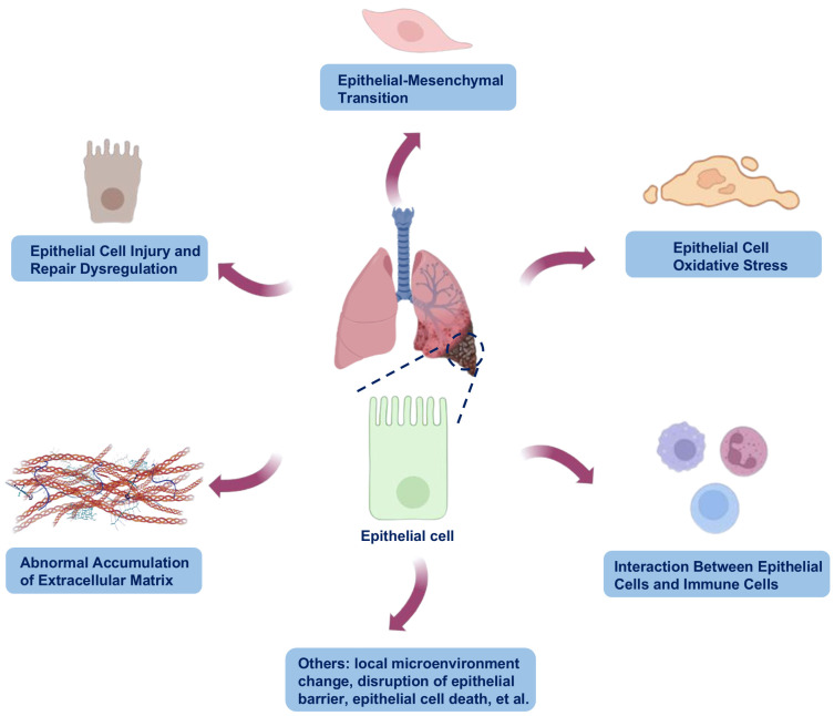
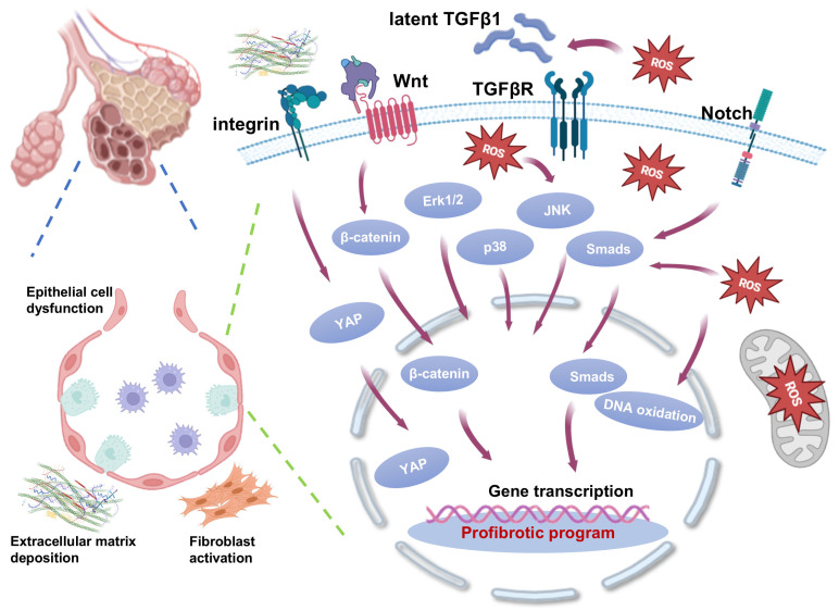
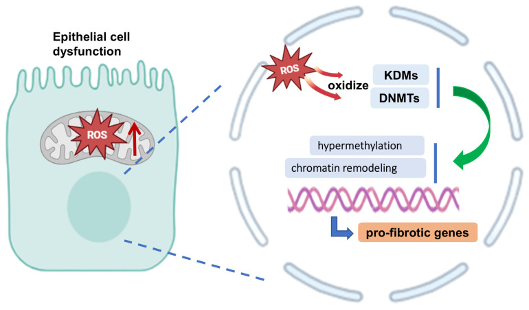
Pulmonary fibrosis (PF) is a progressive and fatal interstitial lung disease characterized by chronic epithelial injury and excessive deposition of extracellular matrix (ECM) driven by dysregulated repair. Increasing evidence has shown that epithelial cell dysfunction plays a key role in PF, involving epithelial-mesenchymal transition (EMT), chronic oxidative stress, disruption of epithelial-immune interactions, and promoting pathological remodeling. Single-cell analyses have identified functionally distinct subpopulations of type 2 alveolar (AT2) cells with pro-fibrotic potential. Epithelial cells exhibit metabolic and epigenetic alterations during PF, which provide new approaches for therapeutic targets. This review summarizes the molecular mechanisms driving epithelial dysfunction in fibrosis progression, with a focus on key regulatory pathways, including transforming growth factor-beta (TGF-β), Wnt, and Notch signaling pathways, as well as miRNA-mediated networks. We also explored emerging epithelial-targeted therapies, ranging from FDA-approved agents (pirfenidone, nintedanib) to experimental inhibitors targeting Galectin-3 and Wnt/β-catenin, providing insights into precision anti-fibrosis strategies for clinical translation.

Keywords: antifibrosis strategies; cell heterogeneity; epithelial dysfunction; epithelial–mesenchymal transition (EMT); pulmonary fibrosis (PF).


Conflict of interest statement

The authors declare no conflicts of interest.

Figures

Figure 1
Figure 1
Possible mechanisms of epithelial involvement in the progression of pulmonary fibrosis (PF). In PF, epithelial cells play multifaceted roles in the fibrotic process. They promote fibrosis through various mechanisms, including epithelial injury, epithelial–mesenchymal transition (EMT), pro-fibrotic factors secretion, extracellular matrix (ECM) remodeling, and inflammatory signaling. Damaged epithelial cells lose their ability to repair, leading to destruction of the alveolar structure and triggering fibrotic responses. The images illustrated in the figures were adapted from https://app.biorender.com (accessed on 26 March 2025).
Figure 2
Figure 2
Crosstalk between epithelial signaling pathways in pulmonary fibrosis (PF). In the pathogenesis of PF, multiple signaling pathways work together to drive disease progression through complex crosstalk and synergistic regulatory networks, which interact at multiple levels, including the receptor level, intracellular signal nodes, and epigenetic level, and ultimately lead to continuous amplification of pro-fibrotic signals and irreversibility of pathological remodeling through dynamic network regulation. The images illustrated in the figures were adapted from https://app.biorender.com (accessed on 27 March 2025).
Figure 3
Figure 3
The changes in DNA methyltransferases (DNMTs) and histone demethylases (KDMs) affected by ROS affect the function of epithelial cells and affect pulmonary fibrosis (PF). In PF, dysfunctional epithelial cells undergo profound molecular rewiring to adopt pro-fibrotic phenotypes. Mitochondrial dysfunction generates excessive ROS, which oxidize DNMTs and KDMs, leading to locus-specific hypermethylation and chromatin remodeling at pro-fibrotic genes. The images illustrated in the figures were adapted from https://app.biorender.com (accessed on 27 March 2025).

Similar articles

References

    1. Henderson N.C., Rieder F., Wynn T.A. Fibrosis: From mechanisms to medicines. Nature. 2020;587:555–566. doi: 10.1038/s41586-020-2938-9. - DOI - PMC - PubMed
    1. Wynn T.A., Ramalingam T.R. Mechanisms of fibrosis: Therapeutic translation for fibrotic disease. Nat. Med. 2012;18:1028–1040. doi: 10.1038/nm.2807. - DOI - PMC - PubMed
    1. Richeldi L., Collard H.R., Jones M.G. Idiopathic pulmonary fibrosis. Lancet. 2017;389:1941–1952. doi: 10.1016/S0140-6736(17)30866-8. - DOI - PubMed
    1. Moss B.J., Ryter S.W., Rosas I.O. Pathogenic Mechanisms Underlying Idiopathic Pulmonary Fibrosis. Annu. Rev. Pathol. 2022;17:515–546. doi: 10.1146/annurev-pathol-042320-030240. - DOI - PubMed
    1. Liu J., Wang F., Hong Y., Luo F. Bibliometric Analysis of the Pirfenidone and Nintedanib in Interstitial Lung Diseases. Heliyon. 2024;10:e29266. doi: 10.1016/j.heliyon.2024.e29266. - DOI - PMC - PubMed

LinkOut - more resources