

Intraoperative Confocal Laser Endomicroscopy Detects Prostate Cancer at the Single-Cell Level with High Specificity and in Real Time: A Preclinical Proof of Concept
- PMID: 40573237
- PMCID: PMC12196187
- DOI: 10.3390/ph18060841
Intraoperative Confocal Laser Endomicroscopy Detects Prostate Cancer at the Single-Cell Level with High Specificity and in Real Time: A Preclinical Proof of Concept
Abstract
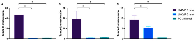
In prostate cancer (PCa) surgery, precise tumor margin identification remains challenging despite advances in surgical techniques. This study evaluates the combination of tumor-specific near-infrared imaging with the PSMA-targeting molecule PSMA-914 and optical endomicroscopy (NIR-pCLE) for single-cell-level tumor identification in a preclinical proof of concept. Methods: NIR-pCLE imaging of varying PSMA-914 concentrations was performed on PSMA-positive LNCaP and PSMA-negative PC-3 cells using Cellvizio® 100 with pCLE Confocal Miniprobes™. To identify optimal PSMA-914 dosing for in vivo imaging, different doses (0-10 nmol) were evaluated using NIR-pCLE, Odyssey CLx imaging, and confocal microscopy in an LNCaP tumor-bearing xenograft model. A proof of concept mimicking a clinical workflow was performed using 5 nmol [68Ga]Ga-PSMA-914 in LNCaP and PC-3 tumor xenografts, including PET/MRI, in/ex vivo NIR-pCLE imaging, and microscopic/macroscopic imaging. Results: NIR-pCLE detected PSMA-specific fluorescence at concentrations above 30 nM in vitro. The optimal dose was identified as 5 nmol PSMA-914 for NIR-pCLE imaging with cellular resolution in LNCaP xenografts. PET/MRI confirmed high tumor uptake and a favorable distribution profile of PSMA-914. NIR-pCLE imaging enabled real-time, single-cell-level detection of PSMA-positive tissue, visualizing tumor heterogeneity, confirmed by ex vivo microscopy and imaging. Conclusions: This preclinical proof of concept demonstrates the potential of intraoperative PSMA-specific NIR-pCLE imaging to visualize tissue structures in real time at cellular resolution. Clinical implementation could provide surgeons with valuable additional information, potentially advancing PCa patient care through improved surgical precision.
Keywords: PSMA; confocal laser endomicroscopy; guided surgery; intraoperative microscopy; prostate cancer.
Conflict of interest statement
A.J., E.M., F.L., A.C. and G.C. are employees of Mauna Kea Technologies. A.-C.E. and M.E. are patent holders on PSMA-targeting inhibitors. All other authors declare no conflicts of interest.
Figures


References
-
- Fernandes S., Williams G., Williams E., Ehrlich K., Stone J., Finlayson N., Bradley M., Thomson R.R., Akram A.R., Dhaliwal K. Solitary pulmonary nodule imaging approaches and the role of optical fibre-based technologies. Eur. Respir. J. 2021;57:2002537. doi: 10.1183/13993003.02537-2020. - DOI - PMC - PubMed
-
- Mauermann J., Fradet V., Lacombe L., Dujardin T., Tiguert R., Tetu B., Fradet Y. The impact of solitary and multiple positive surgical margins on hard clinical end points in 1712 adjuvant treatment-naive pT2-4 N0 radical prostatectomy patients. Eur. Urol. 2013;64:19–25. doi: 10.1016/j.eururo.2012.08.002. - DOI - PubMed
-
- Boorjian S.A., Karnes R.J., Crispen P.L., Carlson R.E., Rangel L.J., Bergstralh E.J., Blute M.L. The impact of positive surgical margins on mortality following radical prostatectomy during the prostate specific antigen era. J. Urol. 2010;183:1003–1009. doi: 10.1016/j.juro.2009.11.039. - DOI - PubMed
Grants and funding
LinkOut - more resources
Full Text Sources
Medical
Miscellaneous
