. 2025 Jun 12;18(6):880.
doi: 10.3390/ph18060880.

Wound-Healing Potential of Myristica fragrans Essential Oil: A Multi-Targeted Approach Involving Inflammation, Oxidative Stress, and Apoptosis Regulation

Affiliations

Wound-Healing Potential of Myristica fragrans Essential Oil: A Multi-Targeted Approach Involving Inflammation, Oxidative Stress, and Apoptosis Regulation

Yahya I Asiri et al. Pharmaceuticals (Basel). .

Abstract

Background: Essential oils are widely studied for their therapeutic potential, including their role in wound healing. Myristica fragrans essential oil (MEO) has been previously investigated for various pharmacological activities, including anti-inflammatory and antimicrobial effects. However, its mechanistic role in accelerating wound healing and modulating critical pathways, such as oxidative stress, inflammation, and apoptosis, remains poorly characterized. MEO contains a rich profile of monoterpene esters, sesquiterpenoids, and phenolic acids, which may contribute to its bioactivity through unique multi-targeted mechanisms. Objective: This research aims to investigate the curative properties of MEO on wound repair, specifically its capacity to regulate inflammation, oxidative stress, and apoptosis in an excision wound model using Wistar rats. Methods: Chemical characterization via GC-MS analysis identified Nitrobenzoate Esters (12.85%), Terpenoid/Cineole (6.99%), and Gamma-Terpinene (4.67%) as the dominant constituents. This study utilized a full-thickness excision wound model, and wound contraction, inflammatory cytokines (IL-1β and TNF-α), a macrophage cell surface marker (CD68), oxidative stress markers (ROS MDA, SOD, GSH), and apoptotic regulation (Caspase-3) was evaluated using macroscopic, histopathological, and immunohistochemical analyses. Result: MEO treatment significantly reduced pro-inflammatory cytokines IL-1β (658.3 ± 32.7 pg/mg, *** p < 0.005) and TNF-α (266.7 ± 33.3 pg/mg, *** p < 0.005), compared to the control group (983.3 ± 60.1 and 650 ± 42.8 ** p < 0.05, respectively). CD68 expression was also markedly decreased (12.67 ± 0.71 ng/mL, *** p < 0.005). Furthermore, MEO effectively attenuated oxidative stress by reducing ROS and MDA levels while restoring antioxidant enzymes GSH and SOD. Conclusions: This study demonstrates that Mace Essential Oil (MEO) effectively promotes wound healing by modulating inflammation, oxidative stress, and apoptosis in a preclinical rat model. Its unique bioactive components suggest significant therapeutic potential as a botanical agent for skin repair. Further research is warranted to explore its application in advanced wound-care formulations.

Keywords: Myristica fragrans; essential oil; mace oil; wound healing.


Conflict of interest statement

The author declares that there are no potential conflicts of interest in this paper.

Figures
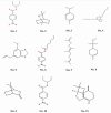
Figure 1
Figure 1
Structure of major compounds identified in an MEO. (No. 1) 4-Nitrobenzoic acid, 3-pentyl ester, (No. 2) Eucalyptol, (No. 3) 1,4-cyclohexadiene, 1-methyl-4-(1-methylethyl), (No. 4) Di-n-octyl phthalate, (No. 5) 1,3-Benzodioxole, 4-methoxy-6-(2-propenyl), (No. 6) 4-Nitro-benzoic acid, 1-methyl-heptyl ester, (No. 7) 2,6-octadien-1-ol, 3,7-dimethyl-, formate, (e)-, (No. 8) 3-cyclohexene-1-methanol, .alpha.,.alpha.,4-trimethyl, (No. 9) Bicyclo[2.2.1]heptane, 7,7-dimethyl-2-methylene-, (No. 10) 4-Nitrobenzoic acid, 3-pentyl ester, and (No. 11) Caryophyllene.
Figure 2
Figure 2
Impact of MEO on wound closure percentage in reference and control groups. n = 6; group, **** p < 0.0001 MEO vs. control, *** p < 0.005 MEO vs. control, ***# p < 0.005 MEO vs. reference group, ** p < 0.05 MEO vs. reference group, * p < 0.05 MEO versus control group, *# p < 0.05. MEO vs. reference group, and *a p < 0.05 reference vs. control group. Tukey’s multiple comparison test was conducted after applying a one-way ANOVA for data analysis. p-values below 0.05 are considered significant.
Figure 3
Figure 3
Representative images showing wound healing among the various groups. (a) The MEO group, (b) reference group, and (c) control group showed signs of wound healing throughout time, as seen by typical photos taken between Days 1 and 21. By the conclusion of the observation period, the MEO treatment group had achieved complete epithelialization and shown accelerated wound healing. Scale bar = 5 mm; applies to all panels. Calibration is based on the known initial wound diameter of 20 mm on Day 1.
Figure 4
Figure 4
Impact of MEO on body weight compared to the reference and control groups. n = 6; mean ± SEM, * p < 0.05 MEO vs. reference, *a p < 0.05 reference vs. control, ** p < 0.05 MEO vs. reference, *** p < 0.005 MEO vs. control, and ***a p < 0.005 MEO vs. reference group; **** p < 0.0001 MEO vs. control group are the corresponding values. Tukey’s multiple comparison test was conducted after the data analysis using a one-way ANOVA. p-values below 0.05 are considered statistically significant.
Figure 5
Figure 5
Impact of MEO on body weight compared to the reference and control groups. n = 6; mean ± SEM, * p < 0.05 MEO vs. reference, *** p < 0.005 MEO vs. control, and ***a p < 0.005 MEO vs. reference group; ***b p < 0.001 reference vs. group and **** p < 0.0001 MEO vs. control group are the corresponding values. Tukey’s multiple comparison test was conducted subsequent to the data analysis using a one-way ANOVA. p-values below 0.05 are considered statistically significant.
Figure 6
Figure 6
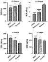
The effects of MEO on ROS, MDA, GSH, and SOD in comparison to the reference and control groups. ** p < 0.05 reference compared to control group, *** p < 0.005 drug compared to control group, * p < 0.05 drug compared to reference group, *# p < 0.05 reference compared to control, and ***# p < 0.005 reference compared to control group, with n = 6 indicating the values, presented as mean ± SEM. Tukey’s multiple comparison test was performed following the one-way ANOVA data analysis. p-values of less than 0.05 are considered statistically significant.
Figure 7
Figure 7
(a) MEO treatment—total restoration of the epithelium (red arrow) and dermis, with minimal infiltrating inflammatory cells (green arrow). (b) Reference: The impacted areas exhibited complete re-epithelialization alongside epidermal hyperplasia (red arrow) and a moderate inflammatory response, characterized by granulation tissue proliferation in the superficial region adjacent to the injury site (green arrow). Additionally, fewer than five blood vessels per high-power field defined moderate angiogenesis or neovascularization (yellow arrow). The region contiguous to the epidermis was substituted. Fibroblasts (blue square) that make collagen and other extracellular matrix components, a network of tiny blood vessels (yellow arrow—capillaries), and different inflammatory cells (green arrow), primarily plasma cells and lymphocytes, followed by neutrophils, are all part of thick granulation tissue. (c) Control: Incomplete wound healing is characterized by inadequate re-epithelialization at the wound site and an overabundance of necrotic debris and inflammatory exudates in the epidermal layers (Red arrow). A considerable inflammatory response with granulation tissue growth was observed in the dermal region around the incision (green arrow), along with pronounced angiogenesis, evidenced by 5 to 10 blood vessels per high-power field (yellow arrow). The area surrounding the epidermal lesion was substituted with granulation tissue, consisting of a network of capillaries (yellow arrow), fibroblasts (blue square) that synthesize collagen and other extracellular matrix components, and various inflammatory cells (green arrow), primarily neutrophils, in addition to plasma cells and lymphocytes.
Figure 8
Figure 8
Immunohistochemical assessments of Caspase-3 activation among each of the groups. (a) MEO-treated group: Caspase-3 expression appeared at normal levels in the fully re-epithelialized epidermal and dermal regions (indicated by red arrows). Normal nucleo-cytoplasmic staining of caspase-3 was evident in the granulation tissue, particularly within fibroblasts (green square) and among infiltrating immune cells, including neutrophils, plasma cells, and lymphocytes (yellow square), located in both the epidermal and dermal layers. (b) Reference group: A moderate level of caspase-3 expression was observed in the healed skin, specifically within the epidermis and dermis (red arrows). Granulation tissue showed moderate nucleo-cytoplasmic caspase-3 localization, primarily in fibroblasts (green square) and immune cells such as neutrophils, plasma cells, and lymphocytes (yellow square), spread across the skin layers. (c) Control group: Caspase-3 expression was limited to a mild level in the re-epithelialized epidermis and dermis (red arrows). Weak nucleo-cytoplasmic caspase-3 staining was detected in fibroblasts (green square) and immune cells (yellow square) within the granulation tissue of both skin layers.

Similar articles

References

    1. Sujatha P.S., Pavithran S., Sujatha P.S. Wound Healing Effect of Furfural and Pentadecanal from Lagerstroemia speciosa (L.) Pers Acetone Flower extracts against Haemadipsa sylvestris Bite. J. Adv. Sci. Res. 2024;15:12–15.
    1. Pereira Beserra F., Sergio Gushiken L.F., Vieira A.J., Augusto Bérgamo D., Luísa Bérgamo P., Oliveira de Souza M., Alberto Hussni C., Kiomi Takahira R., Henrique Nóbrega R., Monteiro Martinez E.R., et al. From inflammation to cutaneous repair: Topical application of lupeol improves skin wound healing in rats by modulating the cytokine levels, NF-κB, Ki-67, growth factor expression, and distribution of collagen fibers. Int. J. Mol. Sci. 2020;21:4952. doi: 10.3390/ijms21144952. - DOI - PMC - PubMed
    1. Ni X., Shan X., Xu L., Yu W., Zhang M., Lei C., Xu N., Lin J., Wang B. Adipose-derived stem cells combined with platelet-rich plasma enhance wound healing in a rat model of full-thickness skin defects. Stem Cell Res. Ther. 2021;12:226. doi: 10.1186/s13287-021-02257-1. - DOI - PMC - PubMed
    1. Shenoy R.R., Sudheendra A.T., Nayak P.G., Paul P., Kutty N.G., Rao C.M. Normal and delayed wound healing is improved by sesamol, an active constituent of Sesamum indicum (L.) in albino rats. J. Ethnopharmacol. 2011;133:608–612. doi: 10.1016/j.jep.2010.10.045. - DOI - PubMed
    1. Kolimi P., Narala S., Nyavanandi D., Youssef A.A. Innovative treatment strategies to accelerate wound healing: Trajectory and recent advancements. Cells. 2022;11:2439. doi: 10.3390/cells11152439. - DOI - PMC - PubMed

LinkOut - more resources