Review
. 2025 May 30;17(6):793.
doi: 10.3390/v17060793.

Insights into the Currently Available Drugs and Investigational Compounds Against RSV with a Focus on Their Drug-Resistance Profiles

Affiliations
Review

Insights into the Currently Available Drugs and Investigational Compounds Against RSV with a Focus on Their Drug-Resistance Profiles

Alessia Magnapera et al. Viruses. .

Abstract

Respiratory syncytial virus (RSV) is a leading cause of severe respiratory illness in infants, young children, as well as elderly and immunocompromised patients worldwide. RSV is classified into two major subtypes, RSV-A and RSV-B, and remains the most frequently detected pathogen in infants hospitalized with acute respiratory infections. Recent advances have brought both passive and active immunization strategies, including FDA-approved vaccines for older adults and pregnant women and new monoclonal antibodies (mAbs) for infant protection. Although significant progress has been made, the need remains for improved antiviral treatments, particularly for vulnerable infants and immunocompromised patients. Recent studies have identified multiple RSV mutations that confer resistance to current treatments. These mutations, detected in both in vitro studies and clinical isolates, often complicate therapeutic outcomes, underscoring the need for updated and effective management strategies. In this context, evaluating protein flexibility through tools like DisoMine provides insight into how specific mutations impact structural dynamics at binding sites, thus affecting ligand affinity. This review aims to synthesize these aspects, offering a comprehensive insight into ongoing efforts to counteract RSV and address the evolving challenge of drug resistance.

Keywords: RSV; drug resistance; mutations; novel drugs; structural dynamics.


Conflict of interest statement

The authors declare no conflicts of interest.

Figures

Figure 1
Figure 1
Schematic representation of the RSV structure and genome. RSV is a negative-sense, single-stranded RNA virus that encodes eleven proteins: nine structural and two non-structural. The non-structural proteins are NS1 and NS2. The viral envelope contains the fusion glycoprotein (F), the attachment glycoprotein (G), and the small hydrophobic protein (SH). The matrix protein (M) is located on the inner surface of the viral envelope. Four nucleocapsid and regulatory proteins act as viral transcription factors: nucleoprotein (N), phosphoprotein (P), large polymerase protein (L), and the M2-1 and M2-2 proteins. Created by Biorender.
Figure 2
Figure 2
RSV replication cycle and target sites for preventive and therapeutic strategies. The figure reports a schematic representation of the RSV replication cycle. The figure also shows an overview of current therapeutics and novel candidates against RSV, including new recombinant antibodies, small molecules such as fusion inhibitors, nucleoprotein inhibitors, nucleoside analogues, and non-nucleoside inhibitors. * Therapeutic strategies not mentioned in this review. F protein (Uniprot P12568), G protein (Uniprot P03423), N protein (Uniprot P03418), L protein (Uniprot P28887), SH protein [72], NS1/NS2 [73]. Created by Biorender.
Figure 3
Figure 3
Cryo-EM structure of the RSV pre-fusion F trimer, derived from the model with PDB ID 8DG9 [93].
Figure 4
Figure 4
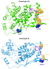
3D monomeric structures of genotype A2, genotype B, and genotype B9320 RSV fusion glycoprotein. In the absence of a resolved 3D structure for genotypes B and B9320, the visualisation of mutations was performed using the PDB-deposited model of genotype A (PDB ID: 8DG9) as a template [79]. Mutated residues are represented in Corey-Pauling-Koltun (CPK); specifically, substitutions in antigenic site A, in F1 domain, in F2 domain, in fusion peptide, in cysteine-rich domain, and in heptad repeat domain are highlighted in yellow, blue, violet, light-blue, red, and green, respectively.
Figure 5
Figure 5
The homodecameric structure, wrapped by the RNA belt, and monomeric structure of the RSV nucleoprotein, derived from the crystallographic model with PDB ID 2WJ8 [126].
Figure 6
Figure 6
3D monomeric structures of genotype A2 and genotype B RSV nucleoprotein. In the absence of a resolved 3D structure for genotypes B, the visualisation of mutations was performed using the PDB-deposited model of genotype A (PDB ID: 2WJ8) as a template [101]. Key amino acid residues for interaction with the phosphoprotein are shown in purple ball-and-sticks. Mutated residues are represented in Corey-Pauling-Koltun (CPK); specifically, those near to the N/P interaction site are shown in yellow, and those more than 10 Angstroms away are shown in blue.
Figure 7
Figure 7
A schematic representation of the RSV G glycoprotein from RSV strain A2. The green residues of the CCD have been resolved in the crystallographic structure with PDB ID 5WNA [109].
Figure 8
Figure 8
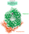
Structure of the RSV polymerase complex (L and P) with a novel non-nucleoside inhibitor (JNJ-8003), derived from the model with PDB ID 8FU3 [144].
Figure 9
Figure 9
3D structure of genotype A2 RSV polymerase, using the model with PDB ID 8FU3 [106]. Mutated residues are represented in Corey-Pauling-Koltun (CPK); specifically, substitutions in motif B are highlighted in yellow, while the M628L substitution in motif F is highlighted in blue.

References

    1. Chanock R., Roizman B., Myers R. Recovery from infants with respiratory illness of a virus related to Chimpanzee Coryza Agent (CCA). I. Isolation, properties and characterization. Am. J. Epidemiol. 1957;66:281–290. doi: 10.1093/oxfordjournals.aje.a119901. - DOI - PubMed
    1. Blount R.E., Jr., Morris J.A., Savage R.E. Recovery of cytopathogenic agent from chimpanzees with coryza. Proc. Soc. Exp. Biol. Med. 1956;92:544–549. doi: 10.3181/00379727-92-22538. - DOI - PubMed
    1. Cantú-Flores K., Rivera-Alfaro G., Carlos Muñoz-Escalante J., Noyola D.E. Global distribution of Respiratory Syncytial Virus A and B infections: A Systematic Review. Pathog. Glob. Health. 2022;116:398–409. doi: 10.1080/20477724.2022.2038053. - DOI - PMC - PubMed
    1. Li Y., Wang X., Blau D.M., Caballero M.T., Feikin D.R., Gill C.J., Madhi S.A., Omer S.B., Simões E.A.F., Campbell H., et al. Global, regional, and national disease burden estimates of acute lower respiratory infections due to respiratory syncytial virus in children younger than 5 years in 2019: A systematic analysis. Lancet. 2022;399:2047–2064. doi: 10.1016/S0140-6736(22)00478-0. - DOI - PMC - PubMed
    1. Munro A.P.S., Martinón-Torres F., Drysdale S.B., Faust S.N. The disease burden of respiratory syncytial virus in Infants. Curr. Opin. Infect. Dis. 2023;36:379–384. doi: 10.1097/QCO.0000000000000952. - DOI - PMC - PubMed

MeSH terms