Case Reports
. 2025 Jun 5;17(6):816.
doi: 10.3390/v17060816.

Case Series of Adverse Pregnancy Outcomes Associated with Oropouche Virus Infection

Affiliations
Case Reports

Case Series of Adverse Pregnancy Outcomes Associated with Oropouche Virus Infection

Daniele Barbosa de Almeida Medeiros et al. Viruses. .

Abstract

The Oropouche virus (OROV) is an arbovirus (Peribunyaviridae: Orthobunyavirus) that traditionally causes febrile outbreaks in Latin America's Amazon region. Previously, OROV was not associated with severe pregnancy outcomes. During the 2022-2024 outbreak in Brazil, OROV expanded geographically, revealing links to adverse pregnancy outcomes. This study describes six cases with varied fetal outcomes, including miscarriage, antepartum, intrauterine fetal demise (IFD), and normal development, correlating with maternal symptoms but not symptom severity. Vertical transmission was confirmed by detecting OROV through RT-qPCR, ELISA, and immunohistochemistry in fetal tissues. Genome sequencing from an IFD case identified a novel reassortment pattern reported in the 2022-2024 outbreak. Severe encephalomalacia, meningoencephalitis, vascular compromise, and multi-organ damage were evident, underscoring the significant risk OROV poses to fetal development and emphasizing the need for further investigation.

Keywords: fetal outcomes; oropouche virus; vertical transmission.


Conflict of interest statement

The authors declare no conflicts of interest.

Figures

Figure 5
Figure 5
Overview of histopathological findings in fetal tissues infected with OROV [9,70,71] compared to ZIKV [57,63,67,68,69]. Both viruses cause severe CNS damage, characterized by intense inflammatory responses, liquefactive necrosis, and neuronal death (necrosis and/or apoptosis). OROV, however, exhibits intense vascular damage and broader systemic involvement.
Figure 1
Figure 1
Flowchart illustrating the timeline of six pregnancy cases, starting from the first day of maternal symptom onset. Maternal information is displayed in blue, including gestational age, symptom duration, and laboratory results confirming OROV infection. Pink shows fetal or newborn data, such as confirmation of fetal death by ultrasound (FDC), date of birth (DOB), sample collection dates, laboratory results, and pregnancy outcomes: miscarriage, antepartum, intrauterine fetal demise (IFD), or normal fetal development (NFD). legend: POS = positive result; RT-qPCR = real-time (RT)-PCR; ELISA = enzyme-linked immunosorbent assay; IHC = immunohistochemistry.
Figure 2
Figure 2
Overview of symptom profiles in pregnant women infected with OROV. Each case is represented by a different color. A wide range of maternal symptoms was observed, regardless of pregnancy outcome. Headache, fever, and myalgia were the most frequently reported symptoms.
Figure 3
Figure 3
Histopathological, and immunohistochemical (IHC) aspects of Intrauterine Fetal Demise, from case 5, associated with OROV and macroscopic findings from case 3. (A,B) Brain: Tissue tearing and fragmentation in the brain, accompanied by activated microglia (circle); presence of a neuron with chromatolysis (black arrow) and congested capillary (red arrow); (C) nervous tissue displaying OROV antigen labeling by IHC in the cytoplasm of neurons (arrow). (D) Fetal necropsy showing epidural hemorrhage in the frontal and occipital regions from case 3. (E,F) Liver: Necrotic hepatocytes (black arrow). Mild inflammatory infiltrate in the portal area (red arrow). Loss of sinusoidal architecture (yellow arrow). (G) Hepatic tissue showing cytoplasmic labeling of hepatocytes (arrows) for the detection of the OROV antigen by IHC. (H,I) Spleen: Hemorrhagic area (black arrow), reactive pattern (circle), and congestion (yellow arrow). (J,K) Kidney: Acute tubular necrosis surrounded by mononuclear cell inflammatory infiltrate (circle); inflammatory infiltrate. (L,M) Heart: Coagulative necrosis with karyolysis in myocardial fibers (black arrow). (N,O) Lung: Detachment of cylindrical cells in small groups (black arrow); mononuclear cell inflammatory infiltrate (circle) and reactive endothelial cell (yellow arrow).
Figure 4
Figure 4
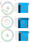
Phylogenetic tree of different OROV strains. Phylogenetic inference was performed using the maximum likelihood (ML) method. (A) ML tree based on the nucleotide (nt) sequences of the S-RNA. The TPM3+I+G4 matrix was used as the best nucleotide substitution model. Measurement of the phylogenetic signal in the dataset showed 31.9% unresolved quartets and 68.1% resolved quartets. The numbers at each major node of the tree correspond to bootstrap values in percentages (1000 replicates). (B) ML tree based on the nt sequences of the M-RNA. The GTR+F+I+R4 matrix was used as the best nucleotide substitution model. Measurement of the phylogenetic signal in the dataset showed 15.2% unresolved quartets and 84.8% resolved quartets. The numbers at each major node of the tree correspond to bootstrap values in percentages (1000 replicates). The scale bar represents nucleotide divergence per site between sequences. The colors represent different countries. (C) ML tree based on the nt sequences of the L-RNA. The GTR+F+I+R2 matrix was used as the best nucleotide substitution model. The measurement of phylogenetic signal in the dataset showed 9.6% unresolved quartets and 90.4% resolved quartets. The numbers at each major node of the tree correspond to bootstrap values in percentage (1000 replicates). The scale bar represents nucleotide divergence per site between sequences. The colors of the sequence IDs represent the countries, and the outer bar represents the decades. The arrow indicates the OROV sequence from fetus BeH891100 (GenBank: S-RNA PQ197204; M-RNA PQ197203; L-RNA PQ197202). Heatmaps showing nucleotide (lower left triangle) and amino acid (upper right triangle) homology identity a matrix between the OROV sequence from the fetus and other OROV sequences from the 2022–2024 outbreak, based on S-RNA (D), M-RNA (E), and L-RNA (F).

References

    1. Souza W.M., Calisher C.H., Carrera J.P., Hughes H.R., Nunes M.R.T., Russell B., Tilson-Lunel N.L., Venter M., Xia H. ICTV Virus Taxonomy Profile: Peribunyaviridae 2024. J. Gen. Virol. 2024;105:002034. doi: 10.1099/jgv.0.002034. - DOI - PMC - PubMed
    1. Chiang J.O., Azevedo R.S., Justino M.C.A., Matos H.J., Cabeça H.L.S., Silva S.P., Henriques D.F., Silva E.V., Andrade G.S., Vasconcelos P.F., et al. Neurological disease caused by Oropouche virus in northern Brazil: Should it be included in the scope of clinical neurological diseases? J. Neurovirol. 2021;27:626–630. doi: 10.1007/s13365-021-00987-9. - DOI - PMC - PubMed
    1. Pinheiro F.P., Freitas A.G., Ohana R.B., Rosa B.A., Travassos da Rosa A.P.A., Juvenal S.L. Meningitis associated with Oropouche virus infections. Rev. Inst. Med. Trop. São Paulo. 1982;24:246–251. - PubMed
    1. Romero-Alvarez D., Escobar L.E. Oropouche fever, an emergent disease from the Americas. Microbes Infect. 2018;20:135–146. doi: 10.1016/j.micinf.2017.11.013. - DOI - PubMed
    1. Pinheiro P., Hoch A.L., Gomes M.L., Roberts D.R. Oropouche virus. IV. Laboratory transmission by Culicoides paraensis. Am. J. Trop. Med. Hyg. 1981;30:172–176. doi: 10.4269/ajtmh.1981.30.172. - DOI - PubMed

Publication types

MeSH terms

Supplementary concepts

Associated data

LinkOut - more resources