Review
. 2025 Jun 6;17(6):817.
doi: 10.3390/v17060817.

The Evolving Role of Zika Virus Envelope Protein in Viral Entry and Pathogenesis

Affiliations
Review

The Evolving Role of Zika Virus Envelope Protein in Viral Entry and Pathogenesis

Ashkan Roozitalab et al. Viruses. .

Abstract

Zika virus (ZIKV) was first discovered in Uganda's Zika Forest in 1947. The early African viruses posed little or no health risk to humans. Since then, ZIKV has undergone extensive genetic evolution and adapted to humans, and it now causes a range of human diseases, including neurologically related diseases in adults and congenital malformations such as microcephaly in newborns. This raises a critical question as to why ZIKV has become pathogenic to humans, and what virological changes have taken place and enabled it to cause these diseases? This review aims to address these questions. Specifically, we focus on the ZIKV envelope (E) protein, which is essential for initiating infection and plays a crucial role in viral entry. We compare various virologic attributes of E protein between the ancestral African strains, which presumably did not cause human diseases, with epidemic strains responsible for current human pathogenesis. First, we review the role of the ZIKV E protein in viral entry and endocytosis during the viral life cycle. We will then examine how the E protein interacts with host immune responses and evades host antiviral responses. Additionally, we will analyze key differences in the sequence, structure, and post-translational modifications between African and Asian lineages, and discuss their potential impacts on viral infection and pathogenesis. Finally, we will evaluate neutralizing antibodies, small molecule inhibitors, and natural compounds that target the E protein. This will provide insights into the development of potential vaccines and antiviral therapies to prevent or treat ZIKV infections and associated diseases.

Keywords: African and Asian lineages; Zika virus; ancestral and epidemic viral strains; antibody-dependent enhancement; envelope protein; inhibitors; microcephaly; neutralizing antibodies; viral entry.


Conflict of interest statement

The authors declare that there is no conflict of interest of any kind. The opinions expressed by the authors contributing to this journal do not necessarily reflect the opinions of the institutions with which the authors are affiliated.

Figures

Figure 1
Figure 1
Schematic representation of the ZIKV viral particle and genome. (A) The Zika viral particle is surrounded by a lipid envelope. The E protein, organized into “rafts” of three E protein dimers lying parallel to each other, covers most of the virion surface. Beneath the E protein lies the M protein. The C protein forms the shell that encloses the viral +ssRNA genome. Phosphatidylserine (PtdSer), a phospholipid, is interlocked with E proteins to form “raft-like” structures on the viral surface. (B) This diagram illustrates the genomic organization of ZIKV, with each viral protein depicted according to its relative position within the RNA genome. Protease cleavage sites are indicated by arrows, representing cleavage by the viral protease, host protease, and furin protease. The numbers above each protein product denote their start and end positions within the genome. The abbreviations used are as follows: anaC (anchored capsid protein C), C (capsid protein C), prM (precursor membrane protein), M (membrane protein), Pr (protein pr), E (envelope protein), NS (nonstructural protein), 2K (signal peptide 2K), and UTR (untranslated region). The viral protease consists of the N-terminal domain of NS3 and the C-terminal domain of NS2B, as described in the text (*). The C-terminal region of NS3 encodes a helicase, while NS5 contains a methyltransferase (MTase) at its N-terminal end and an RNA-dependent RNA polymerase (RdRp) at its C-terminal end. This diagram is adapted from [1].
Figure 2
Figure 2
Schematic overview of the ZIKV life cycle. Upon contact with host cells, ZIKV initiates infection by binding to TAM family receptors on the host cell surface via the bridging molecules Growth arrest-specific 6 (Gas6) and Protein S (ProS), a process mediated by the viral E protein. Note that, besides TAM receptors, other receptors can also be used, which are described in the text. Following attachment, the virus enters host cells through clathrin-mediated endocytosis. Within endosomes, the acidic environment triggers fusion between the viral envelope and the endosomal membrane, leading to the release of the single-stranded, positive-sense viral RNA [ss (+) vRNA] into the cytoplasm. The viral RNA is immediately translated to produce viral proteins. Replication of the viral genome occurs on the ER surface, where double-stranded RNA (dsRNA) intermediates are synthesized and subsequently used to generate additional viral genomes. Immature virions are assembled in the ER, transported through the TGN for maturation, and finally released from the host cell as fully infectious particles. For indications of different color structures on ZIKV, please see Figure 1A.
Figure 3
Figure 3
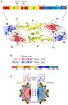
ZIKV E protein and its distinctive structural features between MR766 and BR15 strains. (A) Schematic representation of monomer sequence (top) and 3-D dimer (bottom) of the ZIKV E protein. Structural organization of the E protein monomer, which consists of three N-terminal ectodomains—Domain I (DI), Domain II (DII), and Domain III (DIII)—followed by a C-terminal stem-transmembrane (TM) region that anchors the protein to the viral membrane. Key structural features such as the fusion loop (FL) and glycan loop (GL), along with their respective residue positions, are indicated (bottom). Three-dimensional structure of the E protein dimer, highlighting DI, DII, DIII, and the FL. The 3-D E protein structure is adapted from [44]. (B) Organizational sequence of the E protein monomer shows differences in glycosylation and protonation status between BR15 and MR766. (C) Mirror image comparison of BR15 and MR766 E proteins, highlighting structural differences in the GL. Seven dot connections mark the evolutionarily distinctive a.a. residues within the GL region. The GL shown in the two square boxes illustrates the structural differences between BR15 (red) and MR766 (blue). For indications of different color structures on ZIKV, please see Figure 1A. Abbreviations: Glyco, glycosylation; Prot, protonation state; FL, fusion loop.
Figure 4
Figure 4
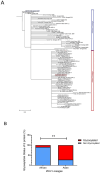
Phylogenetic differences in ZIKV E proteins between African and Asian lineages. (A) Phylogenetic tree analysis of ZIKV E protein sequences derived from available database entries. The tree depicts evolutionary relationships with a specific focus on a.a. residue 156, a key determinant of glycosylation at residue N154. Strains encoding serine (S) or threonine (T) at position 156 are predicted to be glycosylated at N154 [20,56], indicated by a “+” symbol (Gly+), whereas those with isoleucine (I) or other residues at this position are considered non-glycosylated. Two representative strains, BeH8190-BR15 (marked with a red square) and MR766-NIID-Uganda 1947 (marked with a blue square), are highlighted for detailed comparison in the main text. The phylogenetic tree was generated using the neighbor-joining (NJ) and maximum-likelihood (ML) methods, with bootstrap analysis contingent on 1000 replicates based on multiple sequence alignment of ZIKV E proteins using Clustal Omega version 1.2.3. [76]. The unique and complete ZIKV E sequences available in GenBank up to May 2024 were retrieved for this analysis. The resulting trees were visualized using TreeViewer v2.2.0 [77]. The scale bar represents a genetic distance of 0.01, indicating 1% sequence divergence among the aligned E protein sequences. (B) Comparison of the relative abundance of glycosylated (red) versus non-glycosylated (blue) E proteins across lineages. The N-linked glycosylation site at position 154 of the E protein was analyzed among ZIKV strains with unique E protein a.a. sequences, which were obtained from the GenBank virus genome database (http://www.ncbi.nlm.nih.gov/genome/viruses/, accessed on 15 March 2025), Among African lineage viruses, the majority (90.9%; n = 44) lack glycosylation at N154, whereas most Asian lineage viruses (72.3%; n = 36) are glycosylated at this position. Red box, glycosylated; blue box, non-glycosylated. **. Statistical significance, p < 0.005.

References

    1. Lee I., Bos S., Li G., Wang S., Gadea G., Despres P., Zhao R.Y. Probing Molecular Insights into Zika Virus-Host Interactions. Viruses. 2018;10:233. doi: 10.3390/v10050233. - DOI - PMC - PubMed
    1. Dick G.W., Kitchen S.F., Haddow A.J. Zika Virus: A New Virus Isolated from Aedes mosquitoes in Uganda. Nature. 1952;229:735–736.
    1. Bell T.M., Field E.J., Narang H.K. Zika virus infection of the central nervous system of mice. Arch. Gesamte Virusforsch. 1971;35:183–193. doi: 10.1007/BF01249709. - DOI - PubMed
    1. Dick G.W. Zika virus. II. Pathogenicity and physical properties. Trans. R. Soc. Trop. Med. Hyg. 1952;46:521–534. doi: 10.1016/0035-9203(52)90043-6. - DOI - PubMed
    1. Dick G.W., Kitchen S.F., Haddow A.J. Zika virus. I. Isolations and serological specificity. Trans. R. Soc. Trop. Med. Hyg. 1952;46:509–520. doi: 10.1016/0035-9203(52)90042-4. - DOI - PubMed

Publication types

Substances

Associated data

LinkOut - more resources