Development of an SPRi Immune Method for the Quantitative Detection of Osteopontin
- PMID: 40573515
- PMCID: PMC12196602
- DOI: 10.3390/s25123628
Development of an SPRi Immune Method for the Quantitative Detection of Osteopontin
Abstract
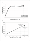
Osteopontin (OPN) is a protein that plays many essential functions in the human body. It is present in most tissues and body fluids. OPN, among other things, participates in wound healing, the formation and remodeling of bone, immune response, inflammation, angiogenesis, and tumor formation. A new analytical method, based on SPRi (surface plasmon resonance imaging) biosensors, has been developed to determine osteopontin in biological fluids. OPN was captured from a solution by an immobilized antibody (mouse or rabbit), a bioreceptor in the SPRi sensor. A separate validation process was carried out for each antibody used. The LOD and LOQ values obtained for the biosensor with mouse antibody were 0.014 ng mL-1 and 0.043 ng mL-1, respectively, and those obtained for the biosensor with rabbit antibody were 0.018 ng mL-1 and 0.055 ng mL-1, respectively. The response ranges of both biosensors were in a similar range: 0.05-1.00 ng mL-1. OPN was determined in blood plasma to demonstrate the sensor potential, showing good agreement with the data obtained using an ELISA test and reported in the literature. The presented method is characterized by ease and speed of measurement, and the process does not require special preparation of samples.
Keywords: ostepontin; surface plasmon resonance imaging (SPRi) biosensor.
Conflict of interest statement
The authors declare no conflicts of interest.
Figures


References
-
- Merritt K. Handbook of Biomaterial Properties. 2nd ed. Springer; New York, NY, USA: 2016. Immune Response; pp. 593–606. - DOI
MeSH terms
Substances
LinkOut - more resources
Full Text Sources
Research Materials

