Review
. 2025 May 22;17(6):682.
doi: 10.3390/pharmaceutics17060682.

Synergistic Cancer Therapies Enhanced by Nanoparticles: Advancing Nanomedicine Through Multimodal Strategies

Affiliations
Review

Synergistic Cancer Therapies Enhanced by Nanoparticles: Advancing Nanomedicine Through Multimodal Strategies

Seyed Mohamad Sadegh Mousavi-Kiasary et al. Pharmaceutics. .

Abstract

Cancer remains a formidable global health challenge due to its complex pathophysiology and resistance to conventional treatments. In recent years, the convergence of nanotechnology and oncology has paved the way for innovative therapeutic platforms that address the limitations of traditional modalities. This review examines how nanoparticle (NP)-based strategies enhance the efficacy of chemotherapy, radiotherapy, phototherapy, immunotherapy, and gene therapy by enabling targeted delivery, controlled drug release, and tumor-specific accumulation via the enhanced permeability and retention (EPR) effect. We discuss the design and functionalization of various organic, inorganic, and hybrid NPs, highlighting their roles in improving pharmacokinetics, overcoming multidrug resistance, and modulating the tumor microenvironment. Particular emphasis is placed on dual and multimodal therapies, such as chemo-phototherapy, chemo-immunotherapy, and gene-radiotherapy, that leverage nanoparticle carriers to amplify synergistic effects, minimize systemic toxicity, and improve clinical outcomes. We also explore cutting-edge advances in gene editing and personalized nanomedicine, as well as emerging strategies to address biological barriers and immunosuppressive mechanisms in the tumor niche. Despite the undeniable promise of nanoparticle-based cancer therapies, challenges related to toxicity, scalable manufacturing, regulatory oversight, and long-term biocompatibility must be overcome before they can fully enter clinical practice. By synthesizing recent findings and identifying key opportunities for innovation, this review provides insight into how nanoscale platforms are propelling the next generation of precision oncology.

Keywords: multimodal treatment; nanoparticles; personalized oncology; synergistic cancer therapy; targeted drug delivery; translational nanomedicine; tumor microenvironment.


Conflict of interest statement

The authors declare no conflict of interest.

Figures

Figure 2
Figure 2
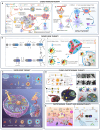
(A) Representation of cancer cell membrane-coated, heat shock protein-functionalized gold nanocages (cmHSP-AuNCs). These nanoparticles demonstrate tumor-targeted delivery and efficient photothermal therapy upon NIR irradiation; “Reprinted from [80], Copyright 2021, with permission from Elsevier” (B) Schematic illustrating the synthesis of sulfur/nitrogen co-doped carbon dots (S, N-CDs), their application for two-photon fluorescence imaging in response to pH, and synergistic photodynamic/photothermal therapy (PDT/PTT) for cancer treatment; “Reprinted (adapted) with permission from [86]. Copyright 2021 American Chemical Society”. (C) Illustration of mannose-functionalized gold nanoparticles (Man@BAu NPs) synthesized via covalent attachment of thiolated mannoside to gold nanoparticles formed in HEPES buffer. Upon exposure to near-infrared (NIR) laser irradiation (808 nm), the Man@BAu NPs exhibit photothermal conversion, elevating local temperature and inducing targeted cancer cell death; “Adapted from [81]”.
Figure 3
Figure 3
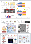
Nanoparticle-based combination cancer therapies integrating multiple conventional treatment modalities. (A) The mechanism and efficacy of PS3D1@DMXAA nanoparticles in breast and melanoma tumor models. Nanoparticles release SN38 and DMXAA within tumor cells via redox-responsive mechanisms, promoting tumor cell death, dendritic cell (DC) maturation, and cytotoxic CD8+ T-cell activation. This approach significantly suppresses primary tumors, reduces lung metastases, and enhances survival by modulating the tumor microenvironment and boosting antitumor immunity; “Reproduced from [115] with permission from, Copyright © 2020, The American Association for the Advancement of Science”. (B) (Bi) Schematic of As-Ap-JNP synthesis via sol-gel methodology, surface functionalization with APTES and succinic anhydride yielding COOH-modified nanoparticles, and final conjugation with aptamer and antisense sequences via chitosan coating; “Reprinted from [116], Copyright 2025, with permission from Elsevier”. (Bii) Preparation of pH-responsive GA-CS-PEI-HBA-DOX@siRNA micelles enabling targeted and controlled co-delivery of doxorubicin (DOX) and siRNA to tumor sites for effective combination therapy; “Reprinted from [117], Copyright 2020, with permission from Elsevier”. (C) Illustration of the hypoxia-responsive RNAi nanomedicine mechanism, designed to silence PGK1 gene expression, thereby sensitizing orthotopic glioblastoma tumors to concurrent chemotherapy and radiotherapy; “Adopted from [118]”. (D) (Di) Design of PDA-coated spiky gold nanoparticles (SGNP@PDA) with TEM image, demonstrating enhanced photothermal stability and synergistic antitumor effects, effectively suppressing primary and distant tumors while establishing sustained immunological protection against tumor recurrence; “Adopted from [119]”. (Dii) Synthesis scheme and mechanism for Pt-HAuNS-PFH@O2 nanoparticles enhancing chemo-photothermal efficacy in breast cancer therapy; “Reprinted from [120], Copyright 2020, with permission from Elsevier”. (Diii) Representation of D/UCNP@cgAuNCs nanoassemblies engineered for tumor microenvironment-responsive ratiometric NIR-II fluorescence imaging and synergistic chemo-photodynamic therapy; “Adopted from [121]”. (E) (Ei) Schematic preparation of AuNP@DCB16F10 nanoparticles, illustrating their mechanism of action for combined photothermal treatment and immune activation; “Adopted from [122]”. (Eii) (a) A schematic shows how gold nanorods (AuNRs) serve as seeds to grow gold nanodendrites (AuNDs), which exhibit a longitudinal LSPR peak in the NIR-II range. (b) Another illustration depicts the use of AuNDs for NIR-II photothermal therapy (PTT), enhancing the effectiveness of cancer immunotherapy; “Reprinted from [123], Copyright 2020, with permission from Elsevier”.
Figure 4
Figure 4
(A) (Ai) (a) Diagram illustrating the synthesis process of Au-DOX@PO-ANG nanoparticles. (b) Schematic showing how Angiopep-2-conjugated, pH-sensitive polymersomes (Au-DOX@PO-ANG) are designed for targeted glioblastoma (GBM) therapy. (c) Biodistribution of Au-DOX@PO-ANG versus free Au-DOX@PO in tumor-bearing mice, with black circles marking tumor regions (ROIs). (d) Bar graph comparing fluorescence intensity in ROIs; ** p < 0.01 indicates statistical significance; “Adopted from [124]”. (Aii) Schematic overview illustrating how radiation influences mitomycin C (MMC) release from Promitil: (a) Promitil passively accumulates in tumors via the enhanced permeability and retention (EPR) effect. (b) Radiation triggers cancer cell death, releasing thiol-based reducing agents. (c) These agents cleave the dithiobenzyl linker in the lipid-based MMC prodrug, enabling controlled drug release at the tumor site; “Reprinted from [125], Copyright 2016, with permission from Elsevier”. (B) Design and evaluation of 10B/siPD-L1 nanoparticles: (a) Schematic shows the synthesis and use of 10B/siPD-L1 nanoparticles for combined boron neutron capture therapy (BNCT) and immunotherapy. (b) ICP-MS spectrum confirms boron isotope content in cRGD-PEG-PME-PBOB. (c) Acid–base titration reveals the buffering behavior of the copolymer. (d) Gel electrophoresis illustrates siRNA binding efficiency at varying 10B-to-siRNA mass ratios. (e) Particle size, morphology (via TEM), and surface charge (zeta potential) are compared before and after nanoparticle crosslinking; “Copyright 2025 Wiley. Used with permission from [126]”. (C) Metastasis suppression using LT-NPs (+L) combined with PD-L1 blockade in a bilateral CT26 tumor model: (a) The treatment schedule involved subcutaneous CT26 tumor implantation followed by intravenous injection to induce lung metastases. Mice received saline, anti-PD-L1, VPF (+L) with anti-PD-L1, or LT-NPs (+L) with anti-PD-L1 on days 0 and 2. Tumors were locally irradiated 6 h post-treatment. (b) Lung images were captured on day 20 to evaluate metastatic burden. (ce) Lung sections were stained with H&E (c), Ki67 and CD8 (d), and TUNEL (e) to assess proliferation, immune infiltration, and apoptosis, respectively. (f) Lung weight was measured as a proxy for tumor load. (g) Survival curves showed treatment outcomes. Statistical significance (ns p > 0.05, * p < 0.05, ** p < 0.01, *** p < 0.001) was analyzed using one-way ANOVA (f) and the log-rank test (g). “Adopted from [127]”. (D) Development of nanoparticles composed of chitosan and hyaluronic acid, facilitating synergistic photothermal and photodynamic therapies through hyperthermia induction and singlet oxygen generation. These nanoparticles stimulate dendritic cell recruitment, immunogenic cell death, T-cell activation, and improve intracellular delivery of therapeutic genes and drugs, augmenting overall anticancer efficacy; “Reprinted from [128], Copyright 2016, with permission from Elsevier.
Figure 5
Figure 5
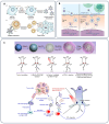
(A) Schematic illustrating the design of engineered cell-membrane-coated nanoparticles for direct antigen presentation: (a) Wild-type tumor cells, which naturally display antigens via MHC-I, are genetically modified to express CD80, a co-stimulatory molecule. Their membranes are then harvested and used to coat polymeric nanoparticle cores. (b) These antigen-presenting nanoparticles (AP-NPs) can directly activate tumor-specific T cells by engaging both the T cell receptor (TCR) and CD28. Once activated, the T cells target and eliminate cancer cells bearing the same antigens, thereby inhibiting tumor growth; “Adapted from [158]”. (B) Schematic of ARAC nanoparticles targeting PD-L1-expressing cancer cells. Initially, ARAC nanoparticles bind PD-L1, are internalized via receptor-mediated endocytosis, and release volasertib to induce G2/M arrest and apoptosis. Surviving cells upregulate PD-L1 expression, which, in turn, enhances subsequent ARAC nanoparticle targeting in a self-amplifying cycle. This ultimately leads to PD-L1 depletion, reactivating cytotoxic CD8+ T cells to mediate antitumor immunity; “Adapted from [136]”. (C) Schematic representation outlining the synthesis of hollow manganese dioxide nanoparticles (H-MnO2-PEG), dual drug loading, and combination therapy with anti-PD-L1 antibodies. Mechanistically, these nanoparticles remodel the tumor microenvironment, promoting polarization of macrophages towards an M1 phenotype and enhancing infiltration of cytotoxic T lymphocytes, thus potentiating anti-PD-L1-mediated immunotherapy; “Adapted from [137]”.
Figure 6
Figure 6
(A) Schematic illustrating the synthesis and application of 2-deoxyglucose-coated PEGylated gold nanoparticles (2DG-PEG-AuD). Upon intravenous administration, nanoparticles preferentially accumulate within tumor tissue, enhancing the efficacy of radiotherapy through localized radiosensitization and improved contrast in computed tomography imaging; “Adapted from [168]”. (B) Illustration depicting intratumorally administered ultrathin bismuth nanosheets (Bi2O2CO3), their intracellular biodegradation, ROS-mediated apoptotic signaling, and resultant enhancement of tumor radiotherapy under low-dose X-ray irradiation; “Copyright 2025 Wiley. Used with permission from [170]”.
Figure 7
Figure 7
(A) (a): Diagram illustrating the synthesis of AuPAMAM nanoparticles using the original pH 6 protocol. (b): TEM images of generation 4 AuPAMAM (AuG4) nanoparticles complexed with GFP plasmid at a 1:30 mass ratio, with DNA visualized using uranyl acetate. Scale bar: 20 nm. (c): Schematic showing how AuPAMAM nanoparticles bind and form complexes with plasmid DNA; “Reprinted from [209], Copyright 2015, with permission from Elsevier”. (B) A schematic representation of a therapeutic strategy for triple-negative breast cancer (TNBC) characterized by c-Myc overexpression, employing CDK1 siRNA delivery via cationic lipid (BHEM-Chol)-facilitated poly(ethylene glycol)-block-poly(d,l-lactide) (PEG5K-PLA11K) nanoparticles; “Reprinted from [188], Copyright 2015, with permission from Elsevier”. (C) (a): Multifunctional siRNA glyconanoparticles (siRNA GlycoNPs) initiate apoptosis in cancer cells. (b): They activate cell death pathways by upregulating receptors like Fas and triggering caspases. Fas detects external death signals, activating caspase-8 and downstream effector caspases. Alternatively, mitochondrial stress can activate caspase-9, ultimately leading to caspase-3 activation and programmed cell death. (c): Additionally, siRNA GlycoNPs silence genes by engaging the RNA interference pathway, causing mRNA degradation or translation inhibition. “Adapted from [197]”.
Figure 1
Figure 1
(A) Schematic depicting polymeric micelles self-assembled from PBA-PEG-SS-PCL-hyd-DOX conjugates. Following intravenous administration, micelles undergo receptor-mediated endocytosis into tumor cells, where intracellular drug release is selectively triggered by elevated glutathione (GSH) levels and acidic intracellular environments; “Reprinted with permission from [63]. Copyright 2025 American Chemical Society”. (B) Schematic illustration highlighting self-assembly of hyaluronic acid-based redox-sensitive micelles (HA-ss-DOCA), their enhanced tumor accumulation via the EPR effect, and subsequent intracellular drug delivery. Internalized micelles undergo receptor-mediated endocytosis, lysosomal escape, and GSH-triggered disassembly to achieve controlled intracellular drug release; “Reprinted from [64] with permission from Elsevier”. (C) Mechanistic illustration of nano drug co-delivery system overcoming multidrug resistance. The nanoparticles selectively release their cargo within tumor cells, bypassing efflux mechanisms mediated by P-gp, and facilitate targeted delivery to nuclear or cytoplasmic therapeutic sites; “Adapted from [65]“. (D) (Di) The diagram contrasts drug release and cellular uptake between targeted and non-targeted PEGylated polymeric nanoparticles. Targeted particles enter cells via receptor-mediated endocytosis. In contrast, non-targeted particles may release drugs into the extracellular space, allowing passive diffusion across the membrane. (Dii) Overview of Paclitaxel and Salinomycin delivery platforms; (a) A schematic of SLM-HA-NP shows how DMAB imparts a positive surface charge, which is partially neutralized by hyaluronic acid. (b) TEM images confirm the spherical morphology and nanoscale size. (c) In vitro release profiles indicate full drug release of both SLM and PTX by day 60. (d) Cytotoxicity evaluation (via MTT assay) after 48 h shows varying toxicity across formulations, including free drugs, encapsulated drugs, targeted systems, and dual-loaded nanoparticles; * p < 0.01 in comparison with SLM, ** p < 0.01 in comparison with SLM + PTX. “Adapted from [66]”.

Similar articles

References

    1. Gavas S., Quazi S., Karpiński T.M. Nanoparticles for Cancer Therapy: Current Progress and Challenges. Nanoscale Res. Lett. 2021;16:173. doi: 10.1186/s11671-021-03628-6. - DOI - PMC - PubMed
    1. Bray F., Laversanne M., Sung H., Ferlay J., Siegel R.L., Soerjomataram I., Jemal A. Global cancer statistics 2022: GLOBOCAN estimates of incidence and mortality worldwide for 36 cancers in 185 countries. CA Cancer J. Clin. 2024;74:229–263. doi: 10.3322/caac.21834. - DOI - PubMed
    1. Siegel R.L., Giaquinto A.N., Jemal A. Cancer statistics, 2024. CA Cancer J. Clin. 2024;74:12–49. doi: 10.3322/caac.21820. - DOI - PubMed
    1. Cooper G., Hausman R. The development and causes of cancer. Cell Mol. Approach. 2000;2:725–766.
    1. Torok J.A., Wu Y., Chino J., Prosnitz L.R., Beaven A.W., Kim G.J., Kelsey C.R. Chemotherapy or combined modality therapy for early-stage Hodgkin lymphoma. Anticancer Res. 2018;38:2875–2881. - PubMed

LinkOut - more resources