

Fabrication of Bio-Composite of Piezoelectric/Myrrh Nanofiber Scaffolds for Wound Healing via Portable Gyrospun
- PMID: 40574028
- PMCID: PMC12196481
- DOI: 10.3390/pharmaceutics17060717
Fabrication of Bio-Composite of Piezoelectric/Myrrh Nanofiber Scaffolds for Wound Healing via Portable Gyrospun
Abstract
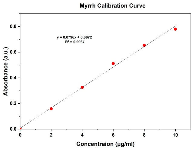
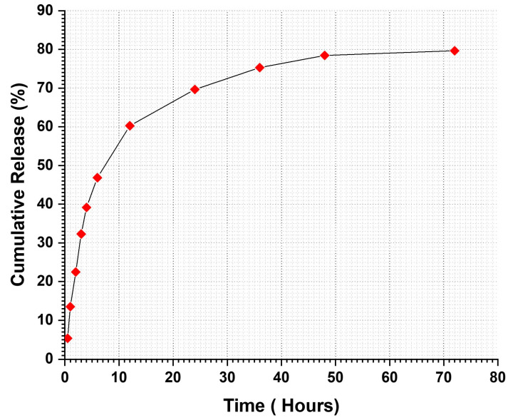
Background/Objectives: Polymeric monoaxial nanofibers are gaining prominence due to their numerous applications, particularly in functional scenarios such as wound management. The study successfully developed and built a special-purpose vessel and device for fabricating polymeric nanofibers. Fabrication of composite scaffolds from piezoelectric poly(vinylidenefluoride-trifluoroethylene) copolymer (PVDF-TrFE) nanofibers encapsulated with myrrh extract was investigated. Methods: The gyrospun nanofibers were characterized using SEM, EDX, FTIR, XRD, and TGA to assess the properties of the composite materials. The study also investigated the release profile of myrrh extract from the nanofibers, demonstrating its potential for sustained drug delivery. The composite's antimicrobial properties were evaluated using the disc diffusion method against various pathogenic microbes, showcasing their effectiveness. Results: It was found that an 18% (w/v) PVDF-TrFE concentration produces the best fiber mats compared to 20% and 25%, resulting in an average fiber diameter of 411 nm. Myrrh extract was added in varying amounts (10%, 15%, and 20%), with the best average fiber diameter identified at 10%, measuring 436 nm. The results indicated that the composite nanofibers were uniform, bead-free, and aligned without myrrh. The study observed a cumulative release of 79.66% myrrh over 72 h. The release profile showed an initial burst release of 46.85% within the first six hours, followed by a sustained release phase. Encapsulation efficiency was 89.8%, with a drug loading efficiency of 30%. Antibacterial activity peaked at 20% myrrh extract. S. mutans was the most sensitive pathogen to myrrh extract. Conclusions: Due to the piezoelectric effect of PVDF-TrFE and the significant antibacterial activity of myrrh, the prepared biohybrid nanofibers will open new avenues toward tissue engineering and wound healing applications.
Keywords: PVDF-TrFE; bio-composite; gyrospun; myrrh; nanofiber; piezoelectric; plant extract; wound healing.
Conflict of interest statement
The authors declare that they have no conflicts of interest.
Figures


Similar articles
-
Drugs for preventing postoperative nausea and vomiting in adults after general anaesthesia: a network meta-analysis.Cochrane Database Syst Rev. 2020 Oct 19;10(10):CD012859. doi: 10.1002/14651858.CD012859.pub2. Cochrane Database Syst Rev. 2020. PMID: 33075160 Free PMC article.
-
Cefadroxil-Mupirocin Integrated Electrospun Nanofiber Films for Burn Wound Therapy.Curr Drug Deliv. 2025 Jun 18. doi: 10.2174/0115672018374558250607134659. Online ahead of print. Curr Drug Deliv. 2025. PMID: 40539348
-
Systemic pharmacological treatments for chronic plaque psoriasis: a network meta-analysis.Cochrane Database Syst Rev. 2021 Apr 19;4(4):CD011535. doi: 10.1002/14651858.CD011535.pub4. Cochrane Database Syst Rev. 2021. Update in: Cochrane Database Syst Rev. 2022 May 23;5:CD011535. doi: 10.1002/14651858.CD011535.pub5. PMID: 33871055 Free PMC article. Updated.
-
Systemic pharmacological treatments for chronic plaque psoriasis: a network meta-analysis.Cochrane Database Syst Rev. 2017 Dec 22;12(12):CD011535. doi: 10.1002/14651858.CD011535.pub2. Cochrane Database Syst Rev. 2017. Update in: Cochrane Database Syst Rev. 2020 Jan 9;1:CD011535. doi: 10.1002/14651858.CD011535.pub3. PMID: 29271481 Free PMC article. Updated.
-
A rapid and systematic review of the clinical effectiveness and cost-effectiveness of debriding agents in treating surgical wounds healing by secondary intention.Health Technol Assess. 2001;5(14):1-131. doi: 10.3310/hta5140. Health Technol Assess. 2001. PMID: 11399237
References
-
- Thomson P. Immunology, microbiology, and the recalcitrant wound. Ostomy/Wound Manag. 2000;46:77S–82S, quiz 83S. - PubMed
LinkOut - more resources
Full Text Sources
