. 2025 Jun 12;17(6):772.
doi: 10.3390/pharmaceutics17060772.

A Natural Latex-Based Smart Dressing for Curcumin Delivery Combined with LED Phototherapy in Diabetic Foot Ulcers: A Pilot Clinical Study

Affiliations

A Natural Latex-Based Smart Dressing for Curcumin Delivery Combined with LED Phototherapy in Diabetic Foot Ulcers: A Pilot Clinical Study

Thamis Fernandes Santana Gomes et al. Pharmaceutics. .

Abstract

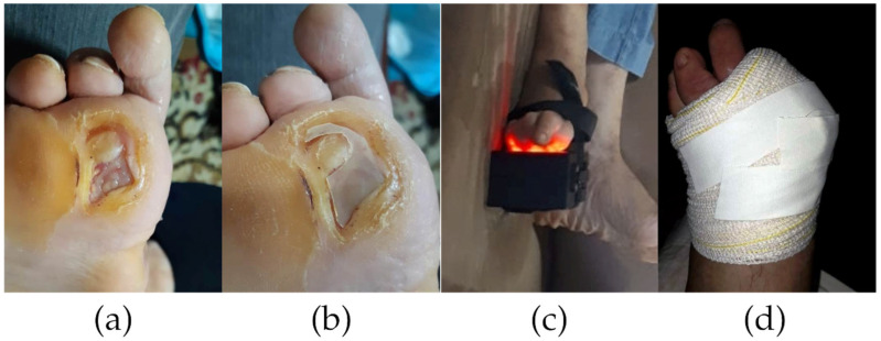
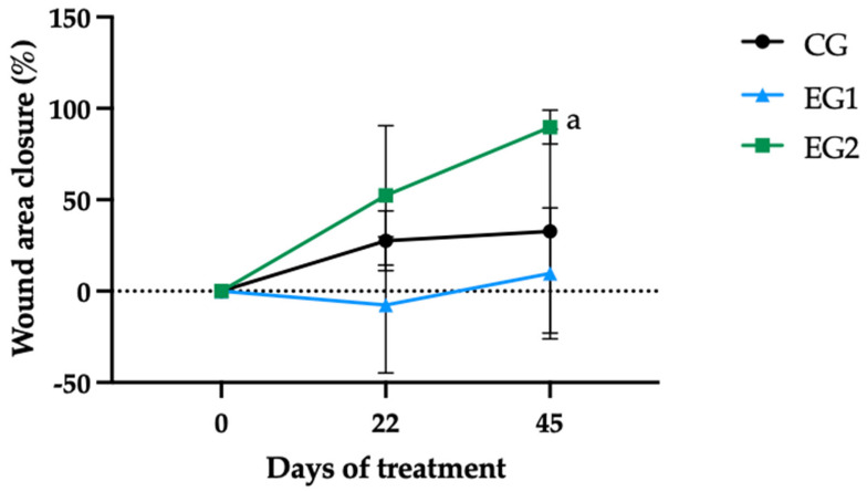
Background: Diabetic foot ulcers (DFUs) affect 25% of diabetes patients, with high risks of amputation (70%), recurrence (65% within 3-5 years), and mortality (50-70% at 5-years). Current treatments are limited by persistent inflammation, oxidative stress, and cost barriers. This study evaluates a bioactive dressing combining a natural latex-based (NLB) biomembrane (Hevea brasiliensis) with curcumin-loaded liposomes, exhibiting angiogenic and antimicrobial properties, and red LED (light-emitting diode) phototherapy (635-640 nm) to address these challenges. Methods: A pilot clinical trial randomized 15 DFU participants into three groups: Control (CG, n = 5, standard care); Experimental Group 1 (EG1, n = 5, NLB + LED, daily treatment); and Experimental Group 2 (EG2, n = 5, NLB-curcumin liposomes + LED, daily treatment). Outcomes included wound closure, inflammatory/oxidative markers, and therapy feasibility. Assessments at D0, D22, and D45 included hematological/biochemical profiling, reactive oxygen species (ROS), and wound area measures. Results: On day 45, GE2 showed an average ulcer contraction of 89.8%, while CG showed 32.8%, and GE1 showed 9.7%. Systemic ROS and biomarkers (C-reactive protein, leukocytes) showed no significant changes (p > 0.05), though transient inflammatory spikes occurred. The combined therapy (EG2) accelerated healing without direct biomarker correlations. Conclusions: These findings highlight the potential of this combined therapy as an accessible, cost-effective DFU treatment, warranting larger studies to optimize home-based protocols and elucidate mechanisms.

Keywords: clinical assay; curcumin; natural latex biomembrane; oxidative stress; ulcers; wound healing.


Conflict of interest statement

The authors declare no conflicts of interest.

Figures

Figure 1
Figure 1
Wound treatment protocol using latex biomembrane and LED phototherapy. (a) Pretreatment presentation of the diabetic ulcer following aseptic cleansing with 0.9% saline solution. (b) Custom-sized natural latex biomembrane being applied to cover the wound bed. (c) Combined therapeutic intervention showing simultaneous biomembrane application and LED phototherapy (635–640 nm wavelength) administration. (d) Completed wound dressing with biomembrane as primary layer, secured by secondary gauze and bandage coverage [35]. Reproduced with permission from Thamis Fernandes Santana, Interdisciplinaridade no contexto das doenças dos pés no diabetes; published by EDUERN, 2021.
Figure 2
Figure 2
Glycated hemoglobin values (%) (a) and blood glucose concentration (b) among the treatment groups: standard therapy (CG), natural latex biomembrane with LED irradiation (EG1), and curcumin-loaded liposome biomembrane with LED irradiation (EG2) at days 0, 22, and 45. Data represent mean values ± standard deviation (n = 5 per group). Statistical significance (p ≤ 0.05) was determined by the Kruskal–Wallis test with post-hoc Dunn for more than two independent samples and Friedman test with post-hoc Dunn for related samples. Superscript letters indicate significant differences compared to day 0.
Figure 3
Figure 3
Leukogram cell counts: (a) leukocytes, (b) segmented neutrophils, (c) eosinophils, (d) basophils, and (e) monocytes among the treatment groups: standard therapy (CG), natural latex biomembrane with LED irradiation (EG1), and curcumin-loaded liposome biomembrane with LED irradiation (EG2) at days 0, 22, and 45. Data represent mean values ± standard deviation (n = 5 per group). Statistical significance (p ≤ 0.05) was determined by the Kruskal–Wallis test with post-hoc Dunn for more than two independent samples and Friedman test with post-hoc Dunn for related samples. The area of the rectangle represents the reference range.
Figure 4
Figure 4
Mean corpuscular volume (a) and mean corpuscular hemoglobin (b) values among the treatment groups: standard therapy (CG), natural latex biomembrane with LED irradiation (EG1), and curcumin-loaded liposome biomembrane with LED irradiation (EG2) at days 0, 22, and 45. Data represent mean values ± standard deviation (n = 5 per group). Statistical significance (p ≤ 0.05) was determined by the Kruskal–Wallis test with post-hoc Dunn for more than two independent samples and Friedman test with post-hoc Dunn for related samples. Superscript letters indicate significant differences compared to the control group. The area of the rectangle represents the reference range.
Figure 5
Figure 5
Concentrations of (a) ultrasensitive C-reactive protein, (b) urea, (c) aspartate aminotransferase, (d) alanine aminotransferase, (e) creatinine, and (f) alkaline phosphatase of the participants among the experimental groups: standard therapy (CG), natural latex biomembrane with LED irradiation (EG1), and curcumin-loaded liposome biomembrane with LED irradiation (EG2) at days 0, 22, and 45. Data represent mean values ± standard deviation (n = 5 per group). Statistical significance (p ≤ 0.05) was determined by the Kruskal–Wallis test with post-hoc Dunn for more than two independent samples and Friedman test with post-hoc Dunn for related samples. The area of the rectangle represents the reference range.
Figure 6
Figure 6
Temporal progression of wound closure (%) across experimental groups: standard therapy (CG), natural latex biomembrane with LED irradiation (EG1), and curcumin-loaded liposome biomembrane with LED irradiation (EG2) at days 0, 22, and 45. Data represent mean values ± standard deviation (n = 5 per group). Statistical significance (p ≤ 0.05) was determined by the Kruskal–Wallis test with post-hoc Dunn for more than two independent samples and Friedman test with post-hoc Dunn for related samples. Superscript letters indicate significant differences compared to day 0. The dashed reference line indicates baseline wound size (0% closure).
Figure 7
Figure 7
Representative wound images and corresponding wound closure percentages (WCP) at baseline (D0), interim (D22), and final evaluation (D45) for all participants across experimental groups: control group (CG, (a)), experimental group 1 (EG1, (b)), and experimental group 2 (EG2, (c)).
Figure 8
Figure 8
Reactive oxygen species (CM•) concentrations in (a) venous blood, (b) wound blood, and (c) wound tissue across treatment groups: standard therapy (CG), natural latex biomembrane with LED irradiation (EG1), and curcumin-loaded liposome biomembrane with LED irradiation (EG2) at days 0, 22, and 45. Data represent mean ± SEM (standard error of mean). Statistical significance (p ≤ 0.05) was determined by the Kruskal–Wallis test with post-hoc Dunn for more than two independent samples and Friedman test with post-hoc Dunn for related samples. Superscript letters indicate significant differences between days 0 and 22 in EG2.

References

    1. Netten J.J., Bus S.A., Apelqvist J., Lipsky B.A., Hinchliffe R.J., Game F., Rayman G., Lazzarini P.A., Forsythe R.O., Peters E.J.G., et al. Definitions and criteria for diabetic foot disease. Diabetes. Metab. Res. Rev. 2020;36:e3268. doi: 10.1002/dmrr.3268. - DOI - PubMed
    1. Jiang X., Zeng Y.-E., Li C., Wang K., Yu D.-G. Enhancing diabetic wound healing: Advances in electrospun scaffolds from pathogenesis to therapeutic applications. Front. Bioeng. Biotechnol. 2024;12:1354286. doi: 10.3389/fbioe.2024.1354286. - DOI - PMC - PubMed
    1. McDermott K., Fang M., Boulton A.J.M., Selvin E., Hicks C.W. Etiology, epidemiology, and disparities in the burden of diabetic foot ulcers. Diabetes Care. 2023;46:209–221. doi: 10.2337/dci22-0043. - DOI - PMC - PubMed
    1. Borderie G., Foussard N., Larroumet A., Blanco L., Barbet-Massin M.-A., Ducos C., Rami-Arab L., Domenge F., Mohammedi K., Ducasse E., et al. The skin autofluorescence of advanced glycation end-products relates to the development of foot ulcers in type 2 diabetes: A longitudinal observational study. J. Diabetes Complicat. 2023;37:108595. doi: 10.1016/j.jdiacomp.2023.108595. - DOI - PubMed
    1. Wang A., Lv G., Cheng X., Ma X., Wang W., Gui J., Hu J., Lu M., Chu G., Chen J., et al. Guidelines on multidisciplinary approaches for the prevention and management of diabetic foot disease (2020 edition) Burn. Trauma. 2020;8:tkaa017. doi: 10.1093/burnst/tkaa017. - DOI - PMC - PubMed

LinkOut - more resources