. 2025 Jun 17;17(6):791.
doi: 10.3390/pharmaceutics17060791.

Exploring a Novel Anti-Inflammatory Therapy for Diabetic Retinopathy Based on Glyco-Zeolitic-Imidazolate Frameworks

Affiliations

Exploring a Novel Anti-Inflammatory Therapy for Diabetic Retinopathy Based on Glyco-Zeolitic-Imidazolate Frameworks

Elena Díaz-Paredes et al. Pharmaceutics. .

Abstract

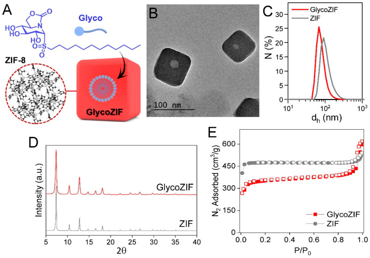
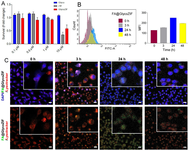
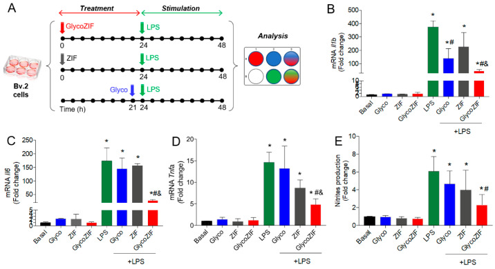
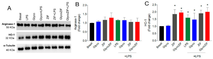
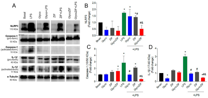
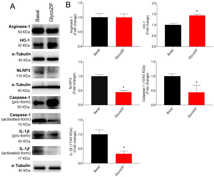
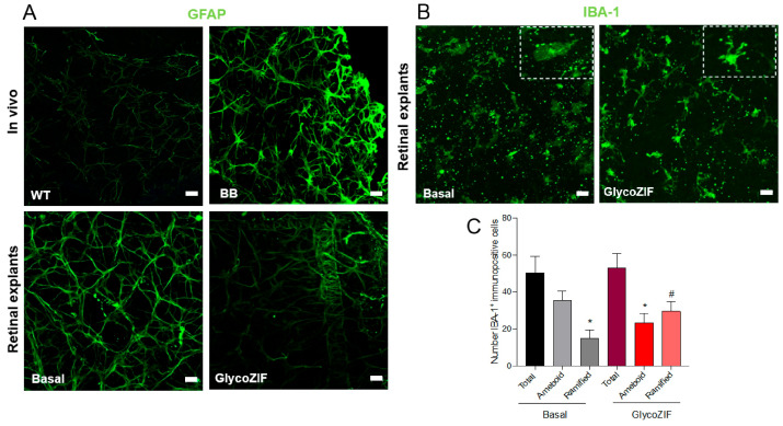
Background/Objectives: Diabetic retinopathy is an ocular disease caused by changes in the expression of inflammatory mediators and increased oxidative stress in the retina and is the leading cause of vision loss in diabetic patients. Currently, there is no treatment capable of reversing retinal damage, which represents a significant burden on the quality of life of patients. (1R)-1-Dodecylsulfonyl-5N,6O-oxomethylidenenojirimycin stands outs as a prototype of the sp2-iminoglycolipids family for its beneficial neuroprotective effect against this chronic eye disease. Critical issues related to the low solubility and bioavailability of this glycolipid in biological settings are overcome by its encapsulation in a Zeolitic-Imidazolate Framework (ZIF) structure, resulting in homogeneous and biocompatible GlycoZIF nanoparticles. Cell studies show an enhanced cellular uptake compared with the free glycolipid, and importantly, its bioactivity is preserved once released inside cells. Methods: Extensive in vitro and ex vivo assays with diabetic retinopathy models unveil the mechanistic pathways of the designed GlycoZIF. Results: A reduction in proinflammatory mediators, increased heme oxygenase-1 level, inhibition of NLRP3 inflammasome, and reduced reactive gliosis is shown. Conclusions: These findings demonstrate for the first time the potential of Glyco-modified ZIFs for the treatment of diabetes-related ocular problems by controlling the immune-mediated inflammatory response.

Keywords: diabetic retinopathy; glycolipid; immune-mediated therapy; inflammation; microglia; zeolitic-imidazolate framework.


Conflict of interest statement

The authors declare no conflicts of interest.

Figures

Figure 1
Figure 1
(A) Chemical structure of the glycolipid studied (Glyco) and scheme of a GlycoZIF particle containing glycolipid molecules encapsulated in the form of micelles. (B) TEM image of the GlycoZIF particles showing the Glyco-micelle inside. (C) DLS number distributions of the GlycoZIF and control ZIF particles dispersed in Milli-Q water. (D) PXRD patterns of the GlycoZIF and ZIF particles. (E) N2 isotherms (77 K) of the GlycoZIF and ZIF particles. Filled symbols represent adsorption, while empty ones represent desorption.
Figure 2
Figure 2
(A) Cell viability of Bv.2 microglial cells under 48 h exposure to increasing concentrations of Glyco, ZIF, or GlycoZIF (from 0.1 to 10 μM) as determined by Crystal Violet assay. Data are presented as fold-change values relative to untreated control cells. (B) Flow cytometric analysis of the uptake of FA@GlycoZIF (500 nM) by Bv.2 cells showing the histograms and the corresponding MFI values as a function of the exposure time (0 h, 3 h, 24 h, and 48 h). (C) Confocal microscopy images of Bv.2 cells incubated with FA@GlycoZIF (500 nM) for different exposure times (0 h, 3 h, 24 h, and 48 h). Green fluorescence corresponds to the encapsulated cargo (i.e., FA), while red fluorescence corresponds to LysoTracker and blue fluorescence to the stained nucleus with DAPI. Higher magnifications (white squares) are shown in the inset top right. Scale bar: 20 μm.
Figure 3
Figure 3
(A) Scheme of the in vitro protocol for optimal treatment with Glyco, ZIF, or GlycoZIF and further LPS stimulation for induction of inflammation in Bv.2 microglial cells. Cells were pretreated with ZIF or GlycoZIF (500 nM) for 24 h or with Glyco (500 nM) for 3 h and then further stimulated with LPS (200 ng/mL) for another 24 h. Quantification of (B) Il1b, (C) Il6, and (D) Tnfa mRNA levels determined by qRT-PCR. (E) Nitrites accumulation determined by the Griess method. Data were normalized to Gapdh gene expression. Results are expressed as mean ± SD (n = 5 independent experiments) and presented as fold-change values relative to untreated control cells (basal value). * p ≤ 0.05 vs. basal treatment, # p ≤ 0.05 vs. LPS treatment, & p ≤ 0.05 vs. Glyco + LPS treatment.
Figure 4
Figure 4
(A) Western blot analyses of proteins extracted from Bv.2 microglial cells subjected to the treatment protocol depicted in Figure 3A. Quantification of (B) Arginase-1 and (C) HO-1 levels. α-Tubulin was used as a loading control. Results are expressed as mean ± SD (n = 4 independent experiments) and presented as fold-change values relative to untreated control cells (basal value). * p ≤ 0.05 vs. basal treatment.
Figure 5
Figure 5
(A) Western blot analyses of proteins extracted from Bv.2 microglial cells subjected to the treatment protocol depicted in Figure 3A. Quantification of (B) NLRP3, (C) Caspase-1, and (D) IL-1β. Activation rate in processed proteins such as caspase-1 and IL-1β is represented as the ratio between activated form and pro-form. α-Tubulin was used as a loading control. Results are expressed as mean ± SD (n = 4 independent experiments) and presented as fold-change values relative to untreated control cells (basal value). * p ≤ 0.05 vs. basal treatment, # p ≤ 0.05 vs. LPS treatment, & p ≤ 0.05 vs. Glyco + LPS treatment.
Figure 6
Figure 6
Ex vivo studies with GlycoZIF nanoparticles. (A) Western blot analyses of proteins extracted from retinal explants of BB rats at 7 weeks old treated with GlycoZIF (500 nM, 24 h). (B) Quantification of the levels of Arginase-1, HO-1, NLRP3, caspase-1, and IL-1β. Activation rate in processed proteins such as caspase-1 and IL-1β is represented as the ratio between the activated form and pro-form. α-Tubulin was used as a loading control. Results are expressed as mean ± SD (n = 4 independent experiments) and presented as fold-change values relative to untreated control cells (basal value). * p ≤ 0.05 vs. basal condition.
Figure 7
Figure 7
Analysis of inflammatory markers in retinas from BB rats during DR progression. (A) GFAP (green) immunostaining in retinal explants from in vivo WT and BB rats or retinal explants at 7 weeks old treated with GlycoZIF (500 nM, 24 h). (B) IBA-1 (green) immunostaining in retinal explants from BB rats at 7 weeks old treated with GlycoZIF (500 nM, 24 h). Scale bar: 20 μm. Zoom images (dashed squares) are shown in the inset top right to clearly see the change in the specific phenotypic shape induced by GlycoZIF treatment. (C) Immunostaining and quantification of IBA-1+ positive cells (ramified or ameboid) in retinal explants from BB rats in basal condition or GlycoZIF treatment. * p ≤ 0.05 vs. IBA-1+ ameboid basal condition, # p ≤ 0.05 vs. IBA-1+ ramified basal condition (n = 5 retinas per condition).

Similar articles

References

    1. Zhang Y., Zhou A. Macrophage activation contributes to diabetic retinopathy. J. Mol. Med. 2024;102:585–597. doi: 10.1007/s00109-024-02437-5. - DOI - PubMed
    1. Tang L., Xu G.T., Zhang J.F. Inflammation in diabetic retinopathy: Possible roles in pathogenesis and potential implications for therapy. Neural Regen. Res. 2023;18:976–982. doi: 10.4103/1673-5374.355743. - DOI - PMC - PubMed
    1. Yue T., Shi Y., Luo S., Weng J., Wu Y., Zheng X. The role of inflammation in immune system of diabetic retinopathy: Molecular mechanisms, pathogenetic role and therapeutic implications. Front. Immunol. 2022;13:1055087. doi: 10.3389/fimmu.2022.1055087. - DOI - PMC - PubMed
    1. Ogurtsova K., Da Rocha Fernandes J.D., Huang Y., Linnenkamp U., Guariguata L., Cho N.H., Cavan D., Shaw J.E., Makaroff L.E. IDF Diabetes Atlas: Global estimates for the prevalence of diabetes for 2015 and 2040. Diabetes Res. Clin. Pract. 2017;128:40–50. doi: 10.1016/j.diabres.2017.03.024. - DOI - PubMed
    1. Saeedi P., Petersohn I., Salpea P., Malanda B., Karuranga S., Unwin N., Colagiuri S., Guariguata L., Motala A.A., Ogurtsova K., et al. Global and regional diabetes prevalence estimates for 2019 and projections for 2030 and 2045: Results from the International Diabetes Federation Diabetes Atlas, 9th edition. Diabetes Res. Clin. Pract. 2019;157:107843. doi: 10.1016/j.diabres.2019.107843. - DOI - PubMed

LinkOut - more resources