

Acquired aberrant partial CD3 expression in recurrent Epstein-Barr virus-negative solitary plasmacytoma of tonsil
- PMID: 40574277
- PMCID: PMC12264490
- DOI: 10.4132/jptm.2025.04.17
Acquired aberrant partial CD3 expression in recurrent Epstein-Barr virus-negative solitary plasmacytoma of tonsil
Abstract
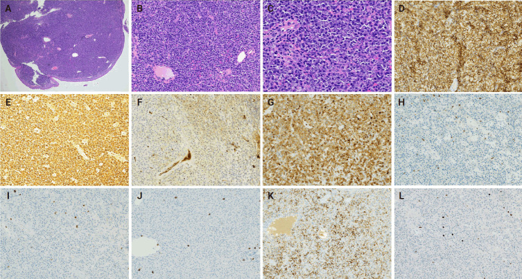
The aberrant expression of specific T-cell maker CD3 in B-cell neoplasms can be a potential diagnostic pitfall leading to a misclassification of cell lineage. Here, we report a case of recurrent solitary plasmacytoma with new aberrant expression of CD3. The neoplastic plasma cells of the recurrent tumor were kappa restricted, positive for CD138, MUM1, negative for CD20, cyclin D1, and Epstein-Barr virus. CD79a was positive in majority of the tumor cells, except for a small focus which was strongly positive for CD3, but negative for other T-cell markers (CD2, CD5, CD7, CD4, and CD8) and CD56. The neoplastic plasma cells of the original tumor were negative for CD3. To the best of our knowledge, only one case of recurrent plasmacytoma with aberrant expression of CD3 has been published, which revealed disease progression in the recurrence. However, we did not observe morphologic evidence of disease progression in our case.
Keywords: CD3; Neoplasm, plasma cell; Plasmacytoma; T-cell antigen.
Conflict of interest statement
The authors declare that they have no potential conflicts of interest to disclose.
Figures


Similar articles
-
Clinical significance of immune cell and biomarker changes in liver cancer.World J Gastrointest Surg. 2025 Jun 27;17(6):104923. doi: 10.4240/wjgs.v17.i6.104923. World J Gastrointest Surg. 2025. PMID: 40584484 Free PMC article.
-
Can a Liquid Biopsy Detect Circulating Tumor DNA With Low-passage Whole-genome Sequencing in Patients With a Sarcoma? A Pilot Evaluation.Clin Orthop Relat Res. 2025 Jan 1;483(1):39-48. doi: 10.1097/CORR.0000000000003161. Epub 2024 Jun 21. Clin Orthop Relat Res. 2025. PMID: 38905450
-
¹⁸F-FDG PET/CT: a review of diagnostic and prognostic features in multiple myeloma and related disorders.Clin Exp Med. 2015 Feb;15(1):1-18. doi: 10.1007/s10238-014-0308-3. Epub 2014 Sep 14. Clin Exp Med. 2015. PMID: 25218739
-
Signs and symptoms to determine if a patient presenting in primary care or hospital outpatient settings has COVID-19.Cochrane Database Syst Rev. 2022 May 20;5(5):CD013665. doi: 10.1002/14651858.CD013665.pub3. Cochrane Database Syst Rev. 2022. PMID: 35593186 Free PMC article.
-
A Case of Idiopathic Follicular Mucinosis Treated Successfully with Cyclosporine.Acta Dermatovenerol Croat. 2024 Dec;32(4):210-211. Acta Dermatovenerol Croat. 2024. PMID: 40657655
References
-
- Lee EJ, Kim M, Kim HS, et al. CD3 and CD20 immunohistochemical staining patterns of bone marrow-infiltrating malignant lymphoma cells. Ann Clin Lab Sci. 2017;47:136–43. - PubMed
-
- Wu B, Vallangeon B, Galeotti J, Sebastian S, Rehder C, Wang E. Epstein-Barr virus-negative diffuse large B cell lymphoma with aberrant expression of CD3 and other T cell-associated antigens: report of three cases with a review of the literature. Ann Hematol. 2016;95:1671–83. doi: 10.1007/s00277-016-2749-0. - DOI - PubMed
LinkOut - more resources
Full Text Sources
Research Materials
Miscellaneous
