

Treatment of recurrent oral carcinoma cuniculatum with immune checkpoint blockade: A case report and literature review
- PMID: 40574746
- PMCID: PMC12199806
- DOI: 10.1016/j.oor.2025.100746
Treatment of recurrent oral carcinoma cuniculatum with immune checkpoint blockade: A case report and literature review
Abstract
Background: Carcinoma cuniculatum (CC) is a rare variant of carcinoma that can arise in the oral cavity. CC can be locally aggressive but rarely metastasizes, and there are few reports of treatment with systemic therapy.
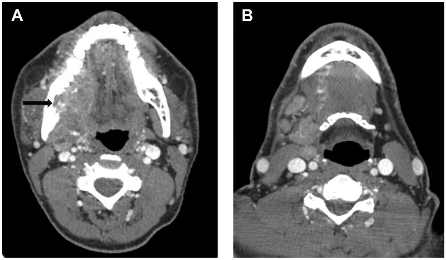
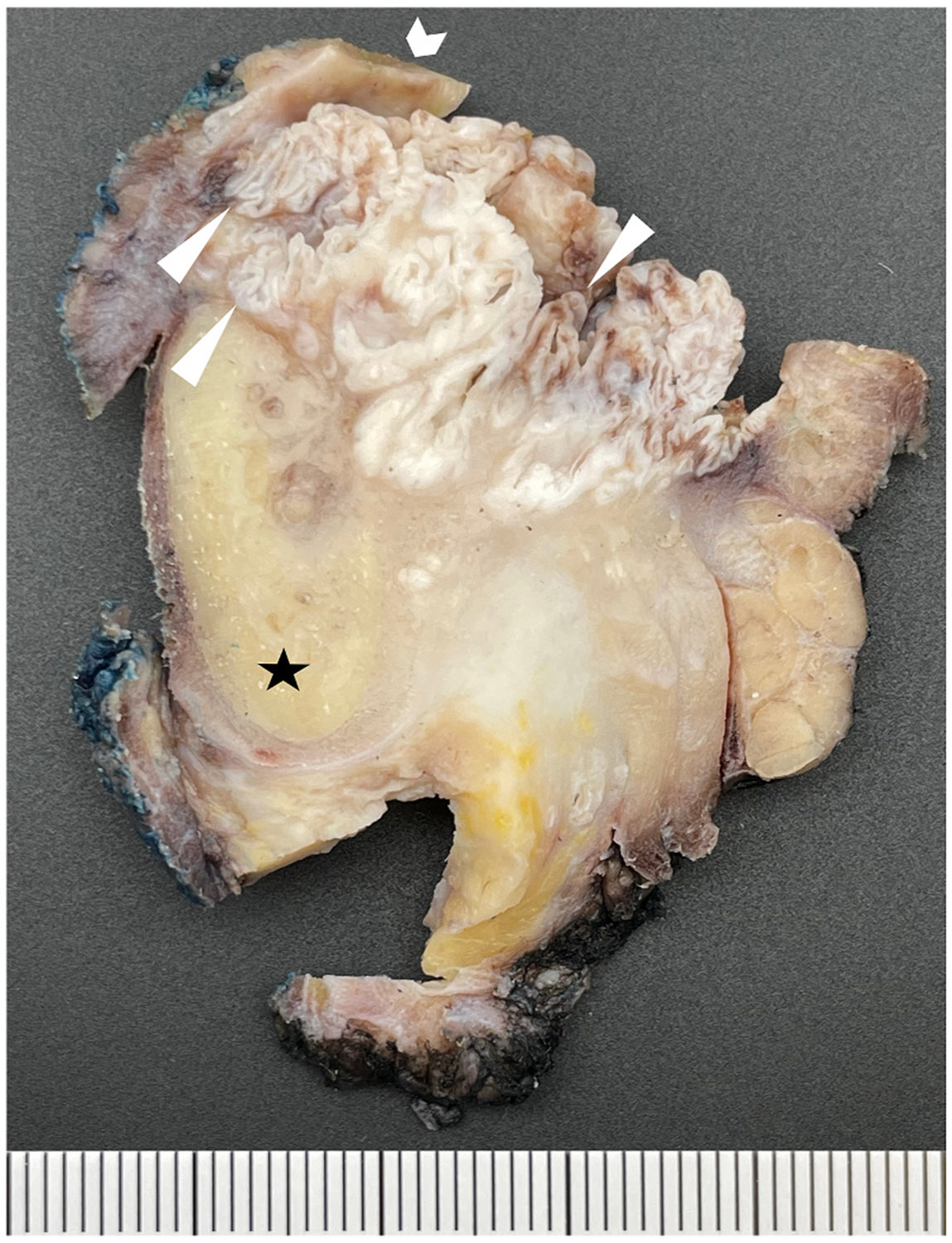
Methods: We present a case of a 44-year-old man with T4bN0 oral carcinoma cuniculatum of the retromolar trigone.
Results: Despite resection with wide margins and adjuvant radiation, the disease recurred 14 months later and was extensive. Following positive margins on a repeat resection, the patient opted for systemic therapy. Despite a PD-L1 combined positive score of 15, the patient did not respond to immunotherapy (pembrolizumab).
Conclusion: Though it has a low propensity for metastasis, oral CC is a locally aggressive variant of squamous cell carcinoma, and recurrences are difficult to treat. Although reports of systemic therapy for CC are limited, this case suggests that immunotherapy may have limited activity in this oral cancer variant.
Keywords: Carcinoma cuniculatum; Head and neck surgery; Immunotherapy; Oral cancer.
Conflict of interest statement
Declaration of competing interest The authors report no conflicts of interest related to this work.
Figures

Similar articles
-
Interventions for preventing oral mucositis in patients with cancer receiving treatment: cytokines and growth factors.Cochrane Database Syst Rev. 2017 Nov 28;11(11):CD011990. doi: 10.1002/14651858.CD011990.pub2. Cochrane Database Syst Rev. 2017. PMID: 29181845 Free PMC article.
-
A rapid and systematic review of the clinical effectiveness and cost-effectiveness of paclitaxel, docetaxel, gemcitabine and vinorelbine in non-small-cell lung cancer.Health Technol Assess. 2001;5(32):1-195. doi: 10.3310/hta5320. Health Technol Assess. 2001. PMID: 12065068
-
Neoadjuvant treatment for stage III and IV cutaneous melanoma.Cochrane Database Syst Rev. 2023 Jan 17;1(1):CD012974. doi: 10.1002/14651858.CD012974.pub2. Cochrane Database Syst Rev. 2023. PMID: 36648215 Free PMC article.
-
EORTC guidelines for the use of erythropoietic proteins in anaemic patients with cancer: 2006 update.Eur J Cancer. 2007 Jan;43(2):258-70. doi: 10.1016/j.ejca.2006.10.014. Epub 2006 Dec 19. Eur J Cancer. 2007. PMID: 17182241
-
Cost-effectiveness of using prognostic information to select women with breast cancer for adjuvant systemic therapy.Health Technol Assess. 2006 Sep;10(34):iii-iv, ix-xi, 1-204. doi: 10.3310/hta10340. Health Technol Assess. 2006. PMID: 16959170
References
-
- Thompson L. World Health Organization classification of tumours: pathology and genetics of head and neck tumours. Ear Nose Throat J 2006;85(2):74. - PubMed
Grants and funding
LinkOut - more resources
Full Text Sources
Research Materials