

Temporal changes in computed tomography findings of a persimmon bezoar: A case report
- PMID: 40574927
- PMCID: PMC11926928
- DOI: 10.12998/wjcc.v13.i18.103426
Temporal changes in computed tomography findings of a persimmon bezoar: A case report
Abstract
Background: Gastric bezoars are masses of indigestible material that accumulate in the stomach, causing nausea, abdominal pain, and vomiting. Persimmon bezoars (diospyrobezoars), which comprise tannins and fibers from persimmons, are relatively rare but may cause significant gastric complications, including gastric outlet obstruction or ileus. Although computed tomography (CT) is a useful imaging tool, diagnosing bezoars can be challenging because their density is similar to that of food debris and gastric content.
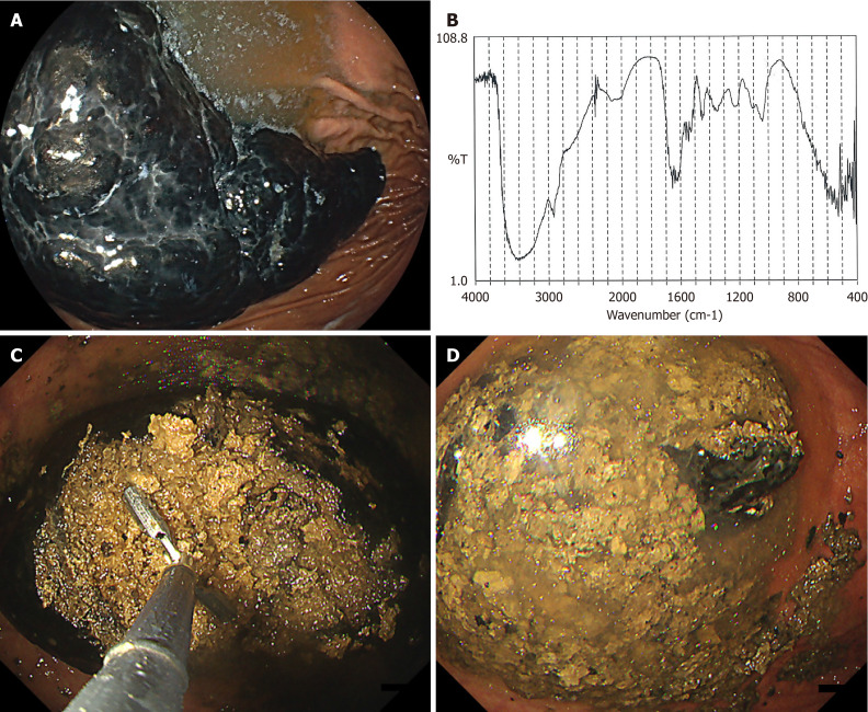
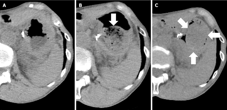
Case summary: Here, we report the case of a 72-year-old woman with a persimmon bezoar that was diagnosed using serial CT imaging and confirmed by endoscopy. CT performed over several months revealed changes in the internal structure and density of the bezoar, suggesting progressive hardening. The patient had a history of a partial gastrectomy and excessive persimmon consumption, both of which are risk factors for bezoar formation. Endoscopic fragmentation of the bezoar successfully resolved symptoms.
Conclusion: Gastric bezoars, particularly persimmon bezoars, present diagnostic challenges because of their variable imaging characteristics. Serial CT can document temporal changes in bezoar density, potentially reflecting changes in hardness. Early diagnosis and endoscopic treatment are essential for effective management, particularly in patients with predisposing factors. This case underscores the importance of considering bezoars in the differential diagnosis of gastric masses, and highlights the value of CT for monitoring changes in bezoar characteristics over time.
Keywords: Case report; Computed tomography; Endoscopic treatment; Gastric bezoar; Persimmon bezoar; Temporal changes.
©The Author(s) 2025. Published by Baishideng Publishing Group Inc. All rights reserved.
Conflict of interest statement
Conflict-of-interest statement: The authors declare no conflict of interest related to this manuscript.
Figures
Similar articles
-
Gastrointestinal phytobezoar following bariatric surgery: Systematic review.Surg Obes Relat Dis. 2016 Nov;12(9):1747-1754. doi: 10.1016/j.soard.2016.09.003. Epub 2016 Sep 9. Surg Obes Relat Dis. 2016. PMID: 27989523
-
Diospyrobezoar (Persimmon Bezoar)-Induced Intestinal Obstruction in an Older Patient: A Case Report.Cureus. 2025 Jul 13;17(7):e87850. doi: 10.7759/cureus.87850. eCollection 2025 Jul. Cureus. 2025. PMID: 40655072 Free PMC article.
-
Signs and symptoms to determine if a patient presenting in primary care or hospital outpatient settings has COVID-19.Cochrane Database Syst Rev. 2022 May 20;5(5):CD013665. doi: 10.1002/14651858.CD013665.pub3. Cochrane Database Syst Rev. 2022. PMID: 35593186 Free PMC article.
-
Surveillance of Barrett's oesophagus: exploring the uncertainty through systematic review, expert workshop and economic modelling.Health Technol Assess. 2006 Mar;10(8):1-142, iii-iv. doi: 10.3310/hta10080. Health Technol Assess. 2006. PMID: 16545207
-
A rapid and systematic review of the clinical effectiveness and cost-effectiveness of topotecan for ovarian cancer.Health Technol Assess. 2001;5(28):1-110. doi: 10.3310/hta5280. Health Technol Assess. 2001. PMID: 11701100
References
-
- Ben-Porat T, Sherf Dagan S, Goldenshluger A, Yuval JB, Elazary R. Gastrointestinal phytobezoar following bariatric surgery: Systematic review. Surg Obes Relat Dis. 2016;12:1747–1754. - PubMed
Publication types
LinkOut - more resources
Full Text Sources
