. 2025 Jun 5;5(6):2491-2499.
doi: 10.1021/jacsau.5c00085. eCollection 2025 Jun 23.

Selective CBP/EP300 Bromodomain Inhibitors: Novel Epigenetic Tools to Counter TNF-α-Driven Inflammation

Affiliations

Selective CBP/EP300 Bromodomain Inhibitors: Novel Epigenetic Tools to Counter TNF-α-Driven Inflammation

Katherine A Gosselé et al. JACS Au. .

Abstract

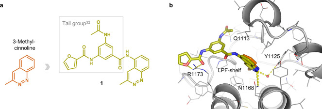
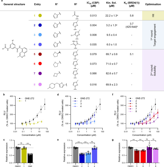
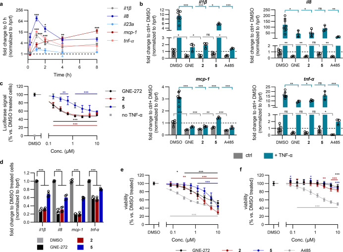
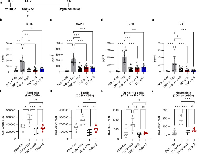
Tumor necrosis factor α (TNF-α) is a central driver of inflammation in autoimmune conditions such as Crohn's disease and rheumatoid arthritis (RA). Targeting epigenetic regulators involved in cytokine expression holds therapeutic promise, yet the precise role of the CBP/EP300 bromodomains (BRDs) in modulating immune responses remains poorly understood. Here, we introduce a distinct class of selective CBP/EP300-BRD inhibitors based on a unique 3-methylcinnoline acetyl-lysine mimic, identified through high-throughput fragment docking. These inhibitors significantly reduce TNF-α-driven cytokine expression in vitro by blocking NFκB signaling in immune cells. In vivo, BRD inhibition led to a robust anti-inflammatory effect, decreasing cytokine secretion (including IL-1β, MCP-1, IL-1α, and IL-6) and preventing immune cell migration to inflamed lymph nodes in a TNF-α-stimulated murine model. Our findings highlight CBP/EP300-BRDs as promising targets for autoimmune therapy, with these non-cytotoxic inhibitors offering a potential complementary approach for RA and other TNF-α-mediated inflammatory conditions.

Keywords: CBP/EP300; NFκB; TNF-α; bromodomain inhibitors; epigenetics; inflammation.


Figures

1
1
Novel acetyl-lysine mimic 3-methylcinnoline, its derived hit compound 1, and their binding modes to the CBP-BRD. (a) Chemical structure of 1a chimera of 3-methylcinnoline and a tail group of the most potent in house developed acetophenone-based inhibitors. (b) Overlay of the co-crystal structure of ligand 1 (olive) in the CBP-BRD binding pocket (PDB code: 6SQM) and the docked 3-methylcinnoline (orange). The key binding interactions are highlighted: direct H-bond interactions with Asn1168 and Arg1173, water mediated interaction with Tyr1125 and stacking interaction with Gln1113. Arg1173 and the Leu-Pro-Phe shelf in CBP/EP300 contribute to the selectivity against BRD4(1).
2
2
Optimization of cinnoline based CBP/EP300-BRD inhibitors. (a) Structural optimization and biochemical data of the inhibitor series; IC50 (CBP) obtained by in-house TR-FRET assay; K D (BRD4(1)) and K D (CBP) by commercial Bromoscan assay; ± st. dev. between technical replicates, a n=4, all other kinetic solubility measurements n=2; bfold-selectivity calculated based on K D (CBP) = 0.009 μM. (b,d,f) Cellular binding of developed inhibitors and GNE-272 to CBP-BRD as determined by the InCELL Pulse assay following 1 h compound treatment of HEK293T cells transiently transfected with ePL-CBP-BRD; RLUrelative light unit. (c,e,g) myc mRNA expression following 4 h treatment of LP1 cells with 1 μM compound; GNE = GNE-272. mRNA expression was quantified by RT-qPCR, and gene expression was normalized to cells treated with DMSO in the same experimental run.
3
3
CBP/EP300-BRD inhibitors target TNF-α-induced inflammation in vitro with reduced toxicity. (a) Cytokine mRNA expression in THP-1 cells following treatment with 10 ng/mL TNF-α. Gene expression was determined by RT-qPCR and normalized to hprt and then to unstimulated cells in the same experimental run. Raw C p values shown in Figure S3. (b) Cytokine mRNA expression following 1 h co-treatment of THP-1 cells with 10 ng/mL TNF-α and 1 μM GNE-272 (abbr. GNE), 2, 5 or A485. mRNA expression quantified by RT-qPCR, normalized to hprt and then to the average of ctrl + DMSO treated cells from all experimental runs. Raw C P values shown in Figure S4. (c) NFκB-RE luciferase reporter assay. HEK293T cells transfected with an NFκB-RE luciferase reporter plasmid were treated for 2 h with BRD inhibitors then stimulated with 10 ng/mL TNF-α for 4 h before luciferase activity was determined. Signal was normalized to cells treated with DMSO and TNF-α on the same plate. (d) Cytokine mRNA expression in THP-1 cells following a therapeutic treatment protocol with BRD inhibitors: 5 h 10 ng/mL TNF-α with 1 μM compounds added 1.5 h after TNF-α stimulation. mRNA expression quantified by RT-qPCR, normalized to hprt and then to cells treated with TNF-α and DMSO in the same experimental run. Raw C p values shown in Figure S6. Cellular viability of (e) THP-1 and (f) MRC5 cells following three-day treatment with compounds. Viability determined using resazurin and normalized to DMSO treated cells on the same plate.
4
4
CBP/EP300-BRD inhibition reduces TNF-α-induced inflammation in vivo. (a) Schematic representation of the experimental set-up. Mice were injected s.c. (footpad) and i.p. with 300 ng of rmTNF-α 90 min before administering s.c. (footpad) and i.p. 10 μL each of the CAPTISOL (abbr. Cap) solutions of 2 (160 ± 1 μM), 5 (790 ± 18 μM) or GNE-272 (715 ± 12 μM) (abbr. GNE). Organs were collected for analysis 5 h post-rmTNF-α administration. Total concentration of IL-1β (b), MCP-1 (c), IL-1α (d), and IL-6 (e), and flow cytometry analysis showing the absolute counts of total lymphocytes (f), T cells (g), dendritic cells (h), and neutrophils (i) in the pLN.

Similar articles

References

    1. Furman D., Campisi J., Verdin E., Carrera-Bastos P., Targ S., Franceschi C., Ferrucci L., Gilroy D. W., Fasano A., Miller G. W., Miller A. H., Mantovani A., Weyand C. M., Barzilai N., Goronzy J. J., Rando T. A., Effros R. B., Lucia A., Kleinstreuer N., Slavich G. M.. Chronic Inflammation in the Etiology of Disease across the Life Span. Nat. Med. 2019;25(12):1822–1832. doi: 10.1038/s41591-019-0675-0. - DOI - PMC - PubMed
    1. Idriss H. T., Naismith J. H.. TNFα and the TNF Receptor Superfamily: Structure-Function Relationship(s) Microsc. Res. Tech. 2000;50(3):184–195. doi: 10.1002/1097-0029(20000801)50:3<184::AID-JEMT2>3.0.CO;2-H. - DOI - PubMed
    1. Esposito E., Cuzzocrea S.. TNF-Alpha as a Therapeutic Target in Inflammatory Diseases, Ischemia- Reperfusion Injury and Trauma. Curr. Med. Chem. 2009;16(24):3152–3167. doi: 10.2174/092986709788803024. - DOI - PubMed
    1. Kany S., Vollrath J. T., Relja B.. Cytokines in Inflammatory Disease. Int. J. Mol. Sci. 2019;20(23):6008. doi: 10.3390/ijms20236008. - DOI - PMC - PubMed
    1. Wajant H., Siegmund D.. TNFR1 and TNFR2 in the Control of the Life and Death Balance of Macrophages. Front. Cell Dev. Biol. 2019;7:91. doi: 10.3389/fcell.2019.00091. - DOI - PMC - PubMed

LinkOut - more resources