Case Reports
. 2025 Jun 12;20(9):4293-4298.
doi: 10.1016/j.radcr.2025.05.060. eCollection 2025 Sep.

Embolization of a high cervical pial arteriovenous fistula resulting in respiratory insufficiency due to diaphragmatic paralysis: A case report

Affiliations
Case Reports

Embolization of a high cervical pial arteriovenous fistula resulting in respiratory insufficiency due to diaphragmatic paralysis: A case report

Jinlu Yu. Radiol Case Rep. .

Abstract

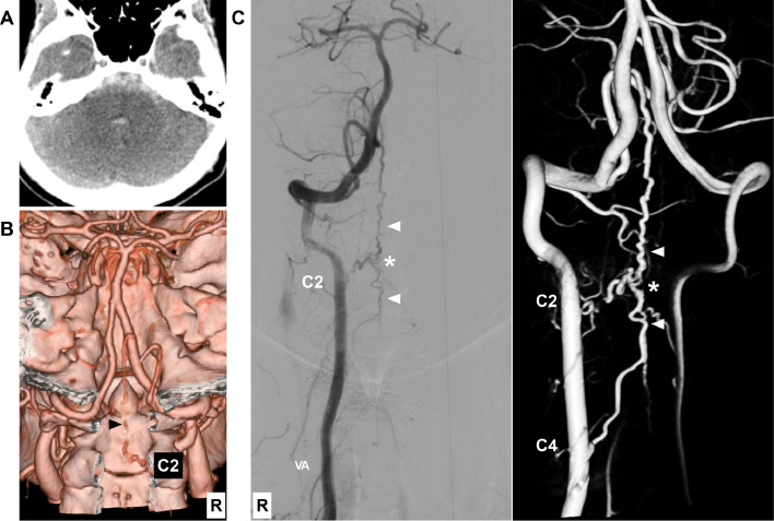
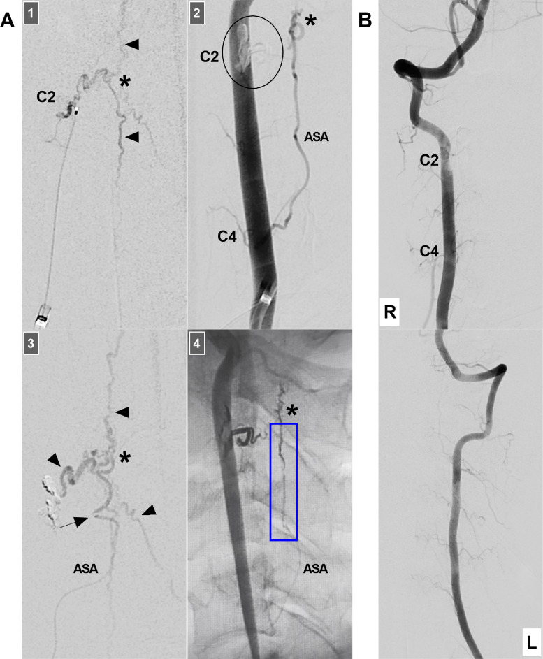
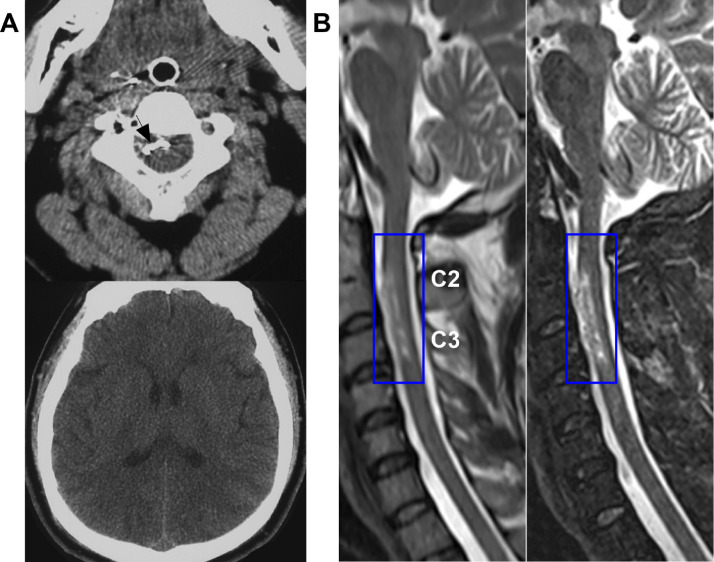
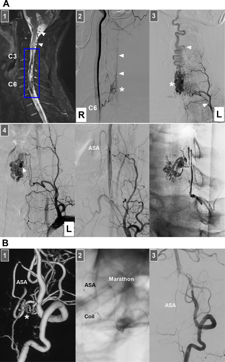
When endovascular treatment (EVT) is administered via the anterior spinal artery (ASA) for high cervical pial arteriovenous fistulas (PAVFs), severe diaphragmatic paralysis, although rare, may occur. We report a 46-year-old male who experienced subarachnoid hemorrhage. The Hunt-Hess scale score was Grade II. Angiography revealed a high cervical PAVF supplied by the C2 radiculomedullary artery and the ASA at the C4 vertebra level. EVT was performed. A microcatheter introduced through the ASA was placed close to the PAVF. After the Onyx agent was cast, the PAVF was obliterated; however, Onyx reflux into the ASA trunk was observed. Postoperatively, the patient had hemiparesis and insufficient autonomous respiration and mechanical ventilation was provided. Fifteen days after EVT, the patient's hemiparesis completely resolved. Ultrasound examination revealed diaphragmatic paralysis. Magnetic resonance imaging revealed cervical cord infarction at the C2-C3 vertebral level, which supported the diagnosis of diaphragmatic paralysis due to phrenic nerve injury from cervical cord infarction above the C4 level. Respiratory function training was performed daily. Eighty-seven days after EVT, his autonomous respiration function further improved. However, 93 days after EVT, the patient died while asleep because the portable ventilator was not used. Therefore, for EVT for high cervical PAVFs, excessive Onyx reflux into the ASA can result in ventral cervical cord infarction, causing severe respiratory insufficiency due to diaphragmatic paralysis. We reported this case to alert neurointerventionists to the lessons learned from experience.

Keywords: Anterior spinal artery; Cervical cord; Diaphragmatic paralysis; Embolization; Pial arteriovenous fistula.


Figures

Fig 1
Fig. 1
Diagnostic images. (A) CT image showing subarachnoid hemorrhage in front of the brainstem and fourth ventricular hemorrhage. (B) CT angiography image showing an abnormal, tortuous vessel extending into the cranium (arrowhead). (C) DSA (left panel) and reconstructive image (right panel) showing a PAVF (asterisks) with cranial and caudal drainages (arrowheads). The feeding arteries derive from the C2 radiculomedullary artery and the ASA from the C4 vertebra level. Abbreviations: ASA, anterior spinal artery; C2 and 4, second and fourth cervical vertebrae; CT, computed tomography; DSA, digital subtraction angiography; PAVF, pial arteriovenous fistula; R, right; VA, vertebral artery.
Fig 2
Fig. 2
Treatment course. (A) Panel 1: Microcatheter angiography via the C2 radiomedullary artery showing the PAVF (asterisk) and its venous drainages (arrowheads); Panel 2: DSA showing that the ASA from the C4 vertebra still feeds the PAVF (asterisk) after the occlusion of the origin of the C2 radiculomedullary artery by casting Onyx (circle); Panel 3: Microcatheter angiography showing the microcatheter tip (arrow) in the ASA close to but not accessing the PAVF (asterisk), with the arrowheads indicating venous drainages; Panel 4: X-ray image showing the Onyx casting within the PAVF (asterisk) and Onyx reflux in the distal ASA (frame). (B) Upper panel: DSA of the right VA showing obliteration of the PAVF; however, the ASA cannot be observed. Lower panel: DSA of the left VA showing no sign of a PAVF. Abbreviations: ASA, anterior spinal artery; C2 and 4, second and fourth cervical vertebrae; DSA, digital subtraction angiography; L, left; PAVF, pial arteriovenous fistula; R, right; VA, vertebral artery.
Fig 3
Fig. 3
Postoperative images. (A) CT image on the fourth postoperative day showing the Onyx (arrow) in the spinal canal and the absorption of subarachnoid hemorrhage. (B) T1-weighted (left panel) and T2-weighted (right panel) MR images taken on the 30th postoperative day revealed infarction (frames) of the cervical cord at the C2-C3 level. Abbreviations: C2-C3, second-third cervical vertebrae; CT, computed tomography; MR, magnetic resonance.
Fig 4
Fig. 4
EVTs for 2 high cervical cord AVFs. (A) EVT in a 35-year-old male. Panel 1: T2-weighted MR image showing hemorrhage (frame) of the cervical cord at the C2–C6 vertebra level; arrowheads indicate dilated veins. Panel 2: DSA of the right VA showing a perimedullary AVF (asterisk) supplied by the C6 radiculomedullary artery with caudal venous drainage (arrowheads). Panel 3: DSA of the left thyrocervical trunk (arrow) showing the ascending cervical artery supplying the AVF (asterisk) with caudal, tortuous venous drainage (arrowhead). Panel 4: Left panel: DSA of the left thyrocervical trunk showing aneurysmal dilation (arrow) in the AVF; Middle panel: DSA showing that the AVF was obliterated by Onyx cast via the ascending cervical artery; Right panel: X-ray image showing the Onyx agent in the AVF and feeding artery. Preoperatively and postoperatively, the patient did not experience respiratory dysfunction. (B) EVT in a 49-year-old male: Panel 1: DSA image showing an AVF ( asterisk) supplied by the ASA; Panel 2: X-ray image showing a detached coil in the distal ASA by the delivery of a Marathon microcatheter; Panel 3: Postoperative DSA showing obliteration of the AVF and preservation of the ASA trunk. Postoperatively, the patient experienced hemiparesis, but 1 month later, he recovered well. Abbreviations: AVF, arteriovenous fistula; ASA, anterior spinal artery; C2-C6, second‒sixth cervical vertebra; DSA, digital subtraction angiography; EVT, endovascular treatment; L, left; MR, magnetic resonance; R, right; VA, vertebral artery.

Similar articles

References

    1. Su H., Yu J. Treatment of high cervical arteriovenous fistulas in the craniocervical junction region. Front Neurol. 2023;14 - PMC - PubMed
    1. Ji T., Guo Y., Shi L., Yu J. Study and therapeutic progress on spinal cord perimedullary arteriovenous fistulas. Biomed Rep. 2017;7(3):214–220. - PMC - PubMed
    1. Meng X., Zhang H., Wang Y., Ye M., He C., Du J., et al. Perimedullary arteriovenous fistulas in pediatric patients: clinical, angiographical, and therapeutic experiences in a series of 19 cases. Childs Nerv Syst. 2010;26(7):889–896. - PubMed
    1. Sasamori T., Hida K., Yano S., Asano T., Iwasaki Y. Cervical perimedullary arteriovenous fistula in an infant presenting with subarachnoid hemorrhage–case report. Neurol Med Chir (Tokyo) 2008;48(9):409–413. - PubMed
    1. Charco-Roca L.M., Simón-Polo E., PC Cuesta-Montero. Ultrasound evaluation of diaphragm function in patients with cervical spinal cord injury: case report. Braz J Anesthesiol. 2024;74(1) - PMC - PubMed

Publication types

LinkOut - more resources