. 2025 Jun 12:12:1615628.
doi: 10.3389/fmed.2025.1615628. eCollection 2025.

Dual locking plate fixation, PRP-augmented autologous bone grafting, and bioactive core construction for femoral fracture nonunion: a retrospective study of 52 cases

Affiliations

Dual locking plate fixation, PRP-augmented autologous bone grafting, and bioactive core construction for femoral fracture nonunion: a retrospective study of 52 cases

Zhihao Peng et al. Front Med (Lausanne). .

Abstract

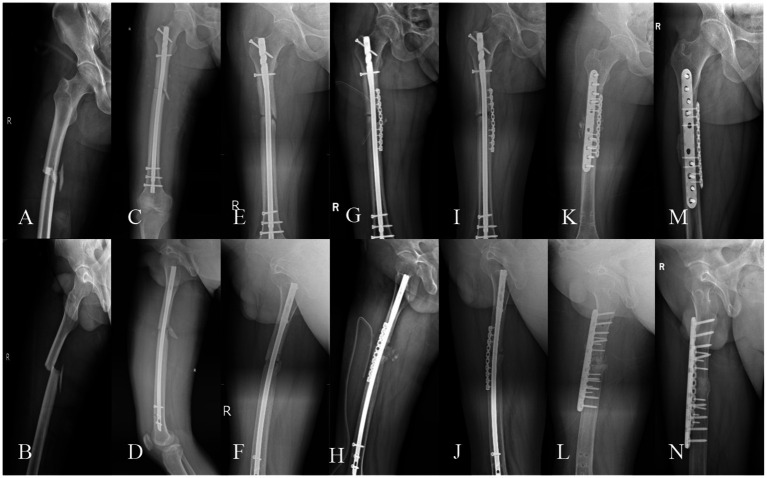
Background: Femoral nonunion remains a challenging orthopedic condition. This study evaluates a combined protocol integrating biomechanical stabilization (dual locking plate fixation) and maximal biological stimulation (PRP-augmented autologous bone grafting with bioactive core construction) to optimize bone healing.

Methods: A retrospective analysis included 52 femoral nonunion patients treated at a tertiary trauma center (2020-2024). Outcomes assessed radiographic union (9-month and final follow-up), clinical union time, thigh incision healing, pain scores (VAS), lower extremity function (LEFS), and complications.

Results: Cohort demographics: 35 males, 17 females; mean age 41.38 years, BMI 24.79 kg/m2. Nonunion subtypes: hypertrophic (36.5%, n = 19), atrophic (50%, n = 26), oligotrophic (13.5%, n = 7); locations: femoral shaft (63.5%, n = 33), supracondylar (36.5%, n = 19). All achieved union (mean follow-up: 19.01 months) with mean union time 6.56 ± 1.04 months. Postoperative outcomes: pain score 0.63 ± 0.97, LEFS 63.92 ± 5.92, incision healing 12.13 ± 1.36 days. The incidence rate of serious complications was 3.85% (2/52).

Conclusion: The protocol demonstrated efficacy and safety, achieving rapid union (6.56 months), robust functional recovery (LEFS 63.92), and a low incidence of serious complications (3.85%). Biomechanical-biological integration represents a viable strategy for femoral nonunion management.

Keywords: autologous iliac bone grafting; dual plate fixation; femoral fracture nonunion; fracture nonunion; platelet-rich plasma.


Conflict of interest statement

The authors declare that the research was conducted in the absence of any commercial or financial relationships that could be construed as a potential conflict of interest.

Figures

Figure 1
Figure 1
(A) Removal of failed internal fixation devices, with nonviable tissues surrounding the fracture ends. (B1) Debridement of nonviable tissues at the fracture site and recanalization of the medullary cavity, revealing the “red pepper sign.” (B2) Cross-sectional view showing fresh, bleeding bone surfaces and a patent, hemorrhagic medullary canal. (C) Mechanical axis correction followed by lateral locking plate placement. (D,E) Harvesting of a corticocancellous bone block from the anterior iliac crest, with additional cancellous bone chips collected. The bone block is trimmed to match the defect geometry. (F) Immersion of the graft in PRP solution. (G) Implantation of PRP-saturated bone grafts into the fracture site, with compression applied via the lateral locking plate and appropriate screw fixation. (H) Placement of an anterior auxiliary plate with compression. (I) Creation of a bioactive chamber by suturing periosteal and residual viable tissues around the fracture site, followed by injection of residual PRP into the chamber and its walls.
Figure 2
Figure 2
(A,B) A full-thickness corticocancellous bone block and cancellous bone chips are harvested from the anterior superior iliac spine region, then contoured to match the defect geometry. (C) Preparation of 10 mL platelet-rich plasma (PRP) at 4–5 × baseline platelet concentration. (D) Immersion of the shaped bone block in PRP. (E) Debridement of nonviable tissues to expose the “red pepper sign” at the fracture ends, followed by mechanical axis correction and deformity reduction. A lateral femoral locking plate is applied. (F) Implantation of the PRP-saturated corticocancellous block into the defect, with cancellous chips packed into residual gaps. (G) An anteromedial locking plate is positioned flush against the grafted bone and compressed.
Figure 3
Figure 3
A 39-year-old male, BMI: 22.16 kg/m2, non-smoker, no metabolic diseases. (A,B) Right femoral shaft fracture sustained in a car accident on January 5, 2021. (C,D) Closed reduction and intramedullary nailing performed on January 13, 2021. (E,F) Nonunion with minimal callus formation observed during follow-up on October 8, 2021. (G,H) Revision surgery on October 23, 2021, retaining the original nail and augmenting with autologous iliac cancellous bone grafting and an auxiliary plate. (I,J) Persistent nonunion noted during follow-up on July 7, 2022. (K,L) Second revision surgery on July 12, 2022, utilizing dual locking plate fixation, PRP-augmented structural iliac bone grafting, and bioactive core construction. (M,N) Excellent radiographic union observed at 6.2 months postoperatively, with a lower extremity functional score of 65 and a VAS pain score of 1.
Figure 4
Figure 4
A 37-year-old male, BMI: 25.98 kg/m2, 2-year smoking history, no metabolic diseases. (A,B) Left distal femoral comminuted fracture sustained in a car accident on January 19, 2022. (C,D) Open reduction and lateral locking plate fixation performed on January 29, 2022. (E,F) Delayed union observed during follow-up on December 23, 2022. (G,H) Nonunion with anterior-medial cortical defect confirmed by CT on June 6, 2023. (I,J) Revision surgery on July 21, 2023, retaining the lateral locking plate and augmenting with an anterior-medial auxiliary plate, PRP-augmented structural iliac bone grafting, and bioactive core construction. (K,L) Radiographic union confirmed at 6.4 months postoperatively (February 2, 2024), with a lower extremity functional score of 60 and a VAS pain score of 0. (M,N) Implant removal performed on March 19, 2024.
Figure 5
Figure 5
A 57-year-old female, BMI: 26.35 kg/m2, sustained a right femoral shaft fracture from a 2-meter fall on March 2, 2024, treated elsewhere with closed reduction and intramedullary nailing. (A,B) Nonunion with minimal callus formation observed during evaluation at our institution on December 15, 2024. (C,D) Revision surgery on December 26, 2024, retaining the original nail and augmenting with an anterior auxiliary plate and PRP-augmented iliac bone grafting. (E,F) Screw cutout at the proximal end of the anterior plate observed during follow-up on February 28, 2025.

Similar articles

References

    1. Wildemann B, Ignatius A, Leung F, Taitsman LA, Smith RM, Pesántez R, et al. Non-union bone fractures. Nat Rev Dis Primers. (2021) 7:57. doi: 10.1038/s41572-021-00289-8, PMID: - DOI - PubMed
    1. Wagner RK, Emmelot MP, Ly TV, Harris MB, Janssen SJ, Kloen P. Long-term patient reported outcomes after revision surgery for lower extremity nonunion: a retrospective cohort study. Injury. (2024) 55:111779. doi: 10.1016/j.injury.2024.111779, PMID: - DOI - PubMed
    1. Nicholson JA, Makaram N, Simpson A, Keating JF. Fracture nonunion in long bones: a literature review of risk factors and surgical management. Injury. (2020) 52:S3–S11. doi: 10.1016/j.injury.2020.11.029 - DOI - PubMed
    1. Siverino C, Metsemakers WJ, Sutter R, Della Bella E, Morgenstern M, Barcik J, et al. Clinical management and innovation in fracture non-union. Expert Opin Biol Ther. (2024) 24:973–91. doi: 10.1080/14712598.2024.2391491, PMID: - DOI - PubMed
    1. Wang C, Sun L, Wang Q, Ma T, Zhang K, Li Z. The technique of “autologous bone grafting through channels” combined with double-plate fixation is effective treatment of femoral nonunion. Int Orthop. (2022) 46:2385–91. doi: 10.1007/s00264-022-05519-6, PMID: - DOI - PubMed

LinkOut - more resources