. 2025 Jun 17;20(1):20221032.
doi: 10.1515/biol-2022-1032. eCollection 2025.

Hesperidin and its zinc(ii) complex enhance osteoblast differentiation and bone formation: In vitro and in vivo evaluations

Affiliations

Hesperidin and its zinc(ii) complex enhance osteoblast differentiation and bone formation: In vitro and in vivo evaluations

Pan Li et al. Open Life Sci. .

Abstract

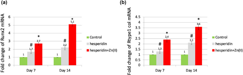
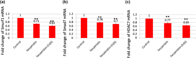
This investigation explores the impact of hesperidin and its zinc(ii) complex on osteoblast differentiation and subsequent bone formation. The biocompatibility of synthesized complexes (0-20 μg/mL) was assessed in vitro using mouse mesenchymal stem cells, while in vivo toxicity was evaluated using a chick embryo model. Both hesperidin and its zinc(ii) complex were found to be non-toxic at a concentration of 10 μg/mL. Notably, these compounds significantly increased alkaline phosphatase activity and enhanced calcium deposition. Molecular analyses revealed upregulation of Runx2 and type 1 collagen mRNA expression, along with increased levels of osteonectin and osteocalcin proteins, while negative regulators of osteoblast differentiation (Smad7, Smurf1, HDAC7) were downregulated. A new aspect of this study is demonstrating that the zinc(ii) complex of hesperidin uniquely enhances osteogenic activity compared to hesperidin alone, highlighting its potential to improve bone formation significantly. Additionally, we elucidated the role of miR-143-3p in mediating these effects, achieved through HDAC7 suppression and enhanced Runx2 expression, assessed using the pmirGLO dual luciferase reporter system. Zebrafish studies further demonstrated the complexes' effects on bone formation, revealing increased osteoblastic activity and improved calcium-to-phosphorus ratios in regenerated scales. These findings underscore the potential of hesperidin-Zn(ii) as a promising therapeutic agent for bone tissue engineering.

Keywords: bone; hesperidin; microRNA; osteoblast; zebrafish; zinc(ii).


Conflict of interest statement

Conflict of interest: Authors state no conflict of interest.

Figures

Figure 1
Figure 1
(a) The MTT assay was conducted to assess the cellular metabolic activity of mouse MSC cells exposed to various concentrations (0–20 μg/mL) of hesperidin and hesperidin–Zn(ii) complexes for up to 48 h. (b) FDA staining was performed to visualize cell morphology and viability after treatment with 10 μg/mL of hesperidin and hesperidin–Zn(ii) complexes for 24 h, followed by observation under a fluorescent microscope. Scale bar: 200 μm. * indicates a statistically significant increase compared to control.
Figure 2
Figure 2
Hesperidin and hesperidin–Zn(ii) complex promoted mouse MSC differentiation toward osteoblasts at the cellular level. (a) Mouse MSCs were treated with different concentrations (5–20 μg/mL) of hesperidin and hesperidin–Zn(ii) complexes for up to 7 days, and ALP activity was measured. (b) Cells treated with 10 μg/mL of hesperidin and hesperidin–Zn(ii) complexes for 7 days were subjected to Alizarin red staining to assess calcium deposition. (c) Quantification of calcium deposition. * indicates a significant increase compared to the control, while # indicates a significant increase compared to hesperidin.
Figure 3
Figure 3
Hesperidin and hesperidin–Zn(ii) complexes increased osteoblast marker gene expression in mouse MSCs. Mouse MSCs were treated with 10 μg/mL of hesperidin and hesperidin–Zn(ii) complexes for up to 14 days. Total RNA was isolated, and the expression of Runx2 mRNA (a) and type 1 collagen mRNA (b) was analyzed by real-time RT-PCR. * indicates a significant increase compared to the control, while # indicates a significant increase compared to hesperidin.
Figure 4
Figure 4
Hesperidin and hesperidin–Zn(ii) complexes increased OC and ON secretion levels in mouse MSCs during osteoblast differentiation. Mouse MSCs were treated with 10 μg/mL of hesperidin and hesperidin–Zn(ii) complexes for up to 14 days. The levels of OC and ON in the conditioned medium were measured. * indicates a significant increase compared to the control, while # indicates a significant increase compared to hesperidin.
Figure 5
Figure 5
Hesperidin and hesperidin–Zn(ii) complexes reduced the expression of negative regulator genes associated with osteoblast differentiation. Mouse MSCs were treated with 10 μg/mL of hesperidin and hesperidin–Zn(ii) complexes for up to 14 days. Total RNA was isolated, and real-time RT-PCR analysis was performed to measure the expression levels of smad7, Smurf1, and HDAC7 mRNAs. ** indicates a significant decrease compared to the control.
Figure 6
Figure 6
Hesperidin and hesperidin–Zn(ii) complexes modulate mir-143 expression in mouse MSCs. Mouse MSCs were treated with 10 µg/mL of hesperidin and hesperidin–Zn(ii) complexes for up to 14 days. (a) Total RNA was isolated, and mir-143 expression was analyzed by real-time RT-PCR. (b) HDAC7 was identified as the predicted target gene for miR-143-3p. (c) Transfection of MG63 cells with miR-143-3p mimic resulted in decreased HDAC7 mRNA and protein expression. (d) Luciferase reporter assay showing direct targeting of HDAC7 3′UTR by miR-143-3p. Wild-type or mutant HDAC7 3′UTR pmirGLO vectors were co-transfected with control miRNA or miR-143-3p mimic in MG63 cells, and luciferase activity was measured after 24 h. ** indicates a significant decrease compared to the control. */# indicates a significant increase compared to the control.
Figure 7
Figure 7
Hesperidin and hesperidin–Zn(ii) complexes enhance bone formation in the zebrafish scale model. Adult zebrafish were treated with 10 µg/mL of hesperidin and hesperidin–Zn(ii) complexes for up to 14 days. (a) Scales were stained with von Kossa staining to visualize calcium deposition. (b) Alizarin red staining was performed to assess mineralization. Quantification of calcium deposition was carried out based on von Kossa staining (c) and Alizarin red staining (d). * indicates a significant increase compared to the control, while # indicates a significant increase compared to hesperidin.
Figure 8
Figure 8
After 14 days of treatment, mineral analysis of the scales was conducted using ICP-MS, and the calcium-to-phosphorus (Ca:P) molar ratio was determined (a). The mRNA expression levels of osteoblast marker genes, including runx2a MASNA isoform, collagen1α, OC, and ON, were analyzed by real-time RT-PCR (b). * denotes a significant increase compared to the control group, while # indicates a significant increase compared to hesperidin treatment.

Similar articles

References

    1. Abdelgawad AAM, El Bassossy TAI, Ahmed FA. Synthesis, chemical characterization, and biological investigation of naturally isolated hesperidin and its metal complexes. Bull Chem Soc Ethiop. 2024;38(2):385–95. 10.4314/bcse.v38i2.8. - DOI
    1. Binkowska I. Hesperidin: synthesis and characterization of bioflavonoid complex. SN Appl Sci. 2020;2:445. 10.1007/s42452-020-2256-8. - DOI
    1. Chen B, Zhu S. Spectroscopic and structural study of the zinc(II)-hesperidin complexes and its analysis application. J Chem Soc Pak. 2018;40(4):682–9.
    1. Chiba H, Uehara M, Wu J, Wang X, Masuyama R, Suzuki K, et al. Hesperidin, a citrus flavonoid, inhibits bone loss and decreases serum and hepatic lipids in ovariectomized mice. J Nutr. 2003;133(6):1892–7. 10.1093/jn/133.6.1892. - DOI - PubMed
    1. Choi SS, Lee SH, Lee KA. A comparative study of hesperetin, hesperidin and hesperidin glucoside: antioxidant, anti-inflammatory, and antibacterial activities in vitro. Antioxidants. 2022;11(8):1618. 10.3390/antiox11081618. - DOI - PMC - PubMed

LinkOut - more resources