

A novel iron bioresorbable scaffold: a potential strategy for pulmonary artery stenosis
- PMID: 40575762
- PMCID: PMC12202099
- DOI: 10.1093/rb/rbaf041
A novel iron bioresorbable scaffold: a potential strategy for pulmonary artery stenosis
Abstract
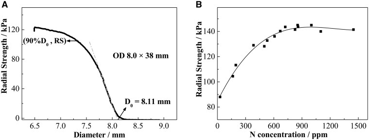
A big diameter bioresorbable scaffold is expected to be used for treatment of vessel stenosis of children with congenital heart disease to adapt the growth characteristics of vessel of children and avoid the late adverse events of permanent stent implanted in children. However, it is challenging to fabricate a big diameter bioresorbable scaffold that is appropriate for percutaneous implantation with enough mechanical performance and can be smoothly delivered in children's small vessel. In this study, a novel iron big and bioresorbable Scaffold (BBS) for pulmonary artery stenosis of children with congenital cardiovascular diseases was fabricated and evaluated. The BBS was made of nitrided iron tube and processed by laser cutting and polishing. The testing results of radial strength, recoil, shortening, maximal expansion diameter and side-branch accessability illustrated the BBS has good mechanical performance. The animal study showed that the percentage of area stenosis of BBSs was 18.1 ± 8.6%, 20.2 ± 5.9% and 20.4 ± 6.1% at 28, 90 and 180 days after implantation in 17 rabbits, and no malposition, thrombus, dissection or tissue necrosis in the rabbit model was detected by micro-CT, STEM and histological examinations. An φ8 × 23 mm BBS was implanted into a 55-month-old child with left pulmonary stenosis, and multiple spiral CT was conducted. No lumen area loss appeared at 1- and 2-year follow-ups in this first-in-man study. It suggested that the BBS might be a new strategy for the therapy of pulmonary artery stenosis in children.
Keywords: big and biodegradable scaffold; congenital heart diseases; intervention; nitrided iron; pulmonary artery stenosis.
© The Author(s) 2025. Published by Oxford University Press.
Figures


Similar articles
-
Home treatment for mental health problems: a systematic review.Health Technol Assess. 2001;5(15):1-139. doi: 10.3310/hta5150. Health Technol Assess. 2001. PMID: 11532236
-
Psychological and/or educational interventions for the prevention of depression in children and adolescents.Cochrane Database Syst Rev. 2004;(1):CD003380. doi: 10.1002/14651858.CD003380.pub2. Cochrane Database Syst Rev. 2004. Update in: Cochrane Database Syst Rev. 2011 Dec 07;(12):CD003380. doi: 10.1002/14651858.CD003380.pub3. PMID: 14974014 Updated.
-
Orthodontic treatment for crowded teeth in children.Cochrane Database Syst Rev. 2021 Dec 31;12(12):CD003453. doi: 10.1002/14651858.CD003453.pub2. Cochrane Database Syst Rev. 2021. PMID: 34970995 Free PMC article.
-
Dexrazoxane for preventing or reducing cardiotoxicity in adults and children with cancer receiving anthracyclines.Cochrane Database Syst Rev. 2022 Sep 27;9(9):CD014638. doi: 10.1002/14651858.CD014638.pub2. Cochrane Database Syst Rev. 2022. PMID: 36162822 Free PMC article.
-
Inhaled magnesium sulfate in the treatment of acute asthma.Cochrane Database Syst Rev. 2017 Nov 28;11(11):CD003898. doi: 10.1002/14651858.CD003898.pub6. Cochrane Database Syst Rev. 2017. PMID: 29182799 Free PMC article.
References
-
- Mainwaring RD, Collins RT, MacMillen KL, Palmon M, Hanley FL. Pulmonary artery reconstruction after failed pulmonary artery stents. Ann Thorac Surg 2020;110:949–55. - PubMed
-
- Myers PO, Tissot C, Beghetti M. Assessment of operability of patients with pulmonary arterial hypertension associated with congenital heart disease. Circ J 2014;78:4–11. - PubMed
-
- Michielon G, Marino B, Formigari R, Gargiulo G, Picchio F, Digilio MC, Anaclerio S, Oricchio G, Sanders SP, Di Donato RM. Genetic syndromes and outcome after surgical correction of tetralogy of fallot. Ann Thorac Surg 2006;81:968–75. - PubMed
-
- Kirklin JW, Blackstone EH, Jonas RA, Shimazaki Y, Kirklin JK, Mayer JE, Pacifico AD, Castaneda AR. Morphologic and surgical determinants of outcome events after repair of tetralogy of fallot and pulmonary stenosis. J Thorac Cardiovasc Surg 1992;103:706–23. - PubMed
-
- Bergersen L, Gauvreau K, Justino H, Nugent A, Rome J, Kreutzer J, Rhodes J, Nykanen D, Zahn E, Latson L, Moore P, Lock J, Jenkins K. Randomized trial of cutting balloon compared with high-pressure angioplasty for the treatment of resistant pulmonary artery stenosis. Circulation 2011;124:2388–96. - PubMed
LinkOut - more resources
Full Text Sources
