Review
. 2025 Sep 1;34(5):440-449.
doi: 10.1097/MNH.0000000000001099. Epub 2025 Jul 3.

Transport and thiazide-inhibition mechanisms of the Na-Cl cotransporter: a structural perspective

Affiliations
Review

Transport and thiazide-inhibition mechanisms of the Na-Cl cotransporter: a structural perspective

Chien-Ling Lee et al. Curr Opin Nephrol Hypertens. .

Abstract

Purpose of review: The structures of the human sodium-chloride cotransporter (hNCC) and its complex with thiazide diuretics have been determined recently. This review summarizes key structural insights into NCC's transport and inhibition mechanisms.

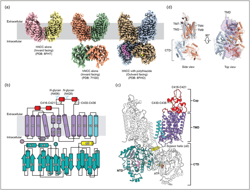
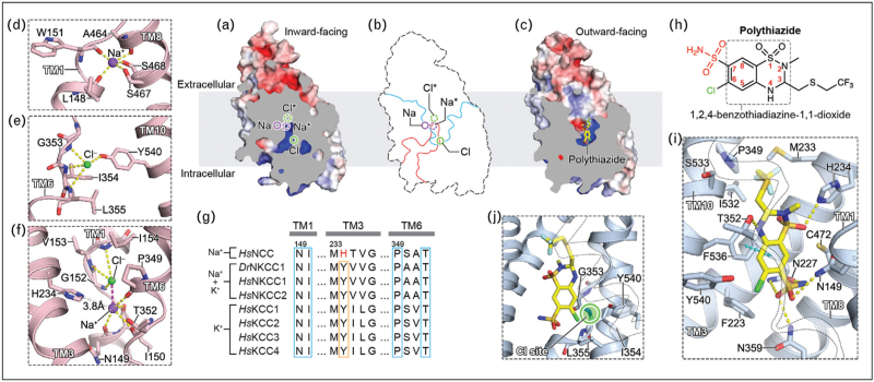
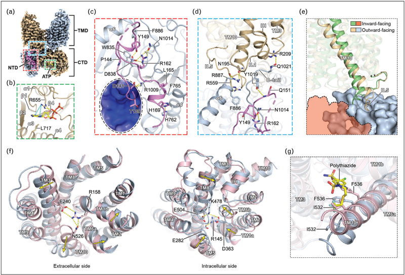
Recent findings: Recent studies revealed the structures of hNCC and its complex with thiazide diuretics, in inward-facing and outward-facing conformations, respectively. The structures of hNCC in two major conformational states provided important insights into the transport and regulatory mechanisms. Thiazide-bound hNCC structures illuminated the molecular mechanisms of thiazide-mediated NCC inhibition and explained the structure-activity relationship of thiazide diuretics.

Summary: Structures of hNCC provide mechanistic insights into molecular mechanisms of loss-of-function NCC variants that cause Gitelman syndrome. The thiazide-bound hNCC structures provide a blueprint for further optimizing thiazide diuretics to reduce side effects. The novel interdomain interaction-mediated hNCC regulatory mechanisms revealed by structural studies lay the foundation for developing next-generation NCC modulators and NCC-rescuing therapeutics for treating NCC dysfunction.

Keywords: cotransporter; cryo-EM; protein structures; sodium-chloride cotransporter; thiazide diuretics; transporter.


Conflict of interest statement

There are no conflicts of interest.

Figures

Box 1
Box 1
no caption available
FIGURE 1
FIGURE 1
Architecture of human sodium-chloride cotransporter. (a) Cryo-EM density maps of hNCC structures. In each structure, the two protomers are colored differently. In the structure of hNCC with polythiazide, the NTD and bound ATP are colored in violet and yellow, respectively. (b) The topology of hNCC. The NTD, TMD, cap domain, and CTD are colored in violet, purple, red, and teal, respectively. TM11 and TM12, which form the interface of the TMD dimer are colored in cyan. The ɑ0 helix (scissor helix) connecting the TMD and CTD is colored in yellow. The two N-glycosylation sites (N406 and N426) and two disulfide bonds (C416-C421 and C430-C436) are labeled. (c) Overall structure of the hNCC dimer. The structure of hNCC in complex with polythiazide (PDB: 8FHO) viewed from the membrane is shown in a ribbon representation. One protomer is highlighted with the coloring scheme the same as Fig. 1b. The bound ATP molecules are shown as yellow sticks. (d) hNCC CTD dimer adopts different orientations in inward- and outward-facing structures. Inward-facing (PDB: 7YG0, orange) and outward-facing (PDB: 8FHO, light blue) hNCC structures are aligned based on the TMD dimer. In the top view, the cross section of the TMD dimer is represented as semitransparent purple areas outlined with dashed lines.
FIGURE 2
FIGURE 2
Substrate- and polythiazide-binding sites. (a) A sliced view of hNCC TMD in an inward-facing conformation. (b) Superimposition of the sliced views of the inward- and outward-facing hNCC TMDs. The outlines of the inward- and outward-facing vestibules are colored in red and blue, respectively. The four ion-binding sites are indicated in Fig. 2a,b. (c) A sliced view of hNCC TMD in an outward-facing conformation. The bound polythiazide is shown as yellow sticks. (d) The Na+-binding “Na site.” In Fig. 2d-f, bound Na+ (purple) and Cl- (green) ions are shown as spheres. Coordinating residues are shown as sticks. Ion-coordinating polar interactions are shown as yellow dashed lines. (e) The Cl--binding “Cl site.” (f) The Na* and Cl* sites. The charge-charge interaction between bound Na+ and Cl- ions is shown as a magenta dashed line. The potential interaction between H234 and the Na+ ion is shown as a white dashed line with the distance labeled. (g) The sequence alignment of the K+(Na*)-binding sites of SLC12 members. Side-chain-coordinating tyrosine residues are highlighted with an orange box. Main-chain-coordinating residues are highlighted with blue boxes. H234 of hNCC is highlighted in red. (h) The structure of polythiazide. The shared scaffold of thiazide diuretics (1,2,4-benzothiadiazine-1,1-dioxide) is outlined with a dashed rectangle and numbered. The 7-position sulfamoyl group and 6-position chlorine are colored in red and green, respectively. (i) The polythiazide-binding site. Polythiazide (yellow) and interacting key residues are shown as sticks. TM6 is made transparent and outlined with dashed lines for clarity. Key polar and π-π interactions between polythiazide and hNCC are shown as yellow and cyan dashed lines, respectively. (j) Polythiazide overlaps with the “Cl site.” TM10 is made transparent and outlined with dashed lines for clarity. The Cl--binding “Cl site” is highlighted with a green circle.
FIGURE 3
FIGURE 3
Regulatory mechanisms and transmembrane domain conformational transitions of human sodium-chloride cotransporter. (a) Cryo-EM density map of hNCC in complex with polythiazide. The protomers with and without a bound NTD are colored in light blue and tan, respectively. The NTD and bound ATP are colored in violet and yellow, respectively. The coloring scheme of Fig. 3b-d is the same as Fig. 3a. The nucleotide-binding site, NTD-CTD interface, and the interface between the NTD-bound CTD and TMD are highlighted with green, red, and blue dashed rectangles, respectively. (b) The nucleotide-binding site. ATP (yellow) and key interacting residues are depicted in stick representation. In Fig. 3b-d and f, key polar and cation-π interactions are shown as yellow and cyan dashed lines, respectively. (c) The NTD-CTD interface. Key residues involved in NTD-CTD interactions are shown as sticks. A key NTD phosphoacceptor, T139 (equivalent to T60 in hNCC), is also shown as sticks. The surface of the CTD close to T139 is shown and colored by electrostatic potential (white, 0 kTe−1; blue, +5 kTe−1). (d) The interface between the NTD-bound CTD and TMD. For the TMD and CTD, regions other than TM3, IL1, TM10, IL5, α6, and C-tail are made semitransparent for clarity. Key residues involved in NTD-CTD and TMD-CTD interactions are shown as sticks. (e) Comparisons of the TMD-CTD interfaces of the inward- and outward-facing hNCC. The TMD-CTD interface of the NTD-bound CTD in the outward-facing structure (PDB: 8FHO) is compared to the corresponding TMD-CTD interface in the inward-facing structure (PDB: 7YG0). The two structures are aligned using the TMDs, which are shown as ribbon representations. The CTDs of the inward- and outward-facing structures are shown as a dashed line-outlined diagram (orange) and a surface representation (light blue), respectively. The TMDs of the inward- and outward-facing structures are colored in green and tan, respectively. The TMDs other than TM10 and IL5 are made semitransparent for clarity. (f) Conformational changes of the TMD from inward-facing (pink) to outward-facing (light blue). The left and right panels are viewed from the extracellular and intracellular sides, respectively. Movements of TM helices undergoing substantial conformational changes are indicated with yellow arrows. Extracellular and intracellular gate residues are shown as sticks. (g) Superposition of the inward-facing (pink) and polythiazide-bound outward-facing (light blue) hNCC structures. Polythiazide is shown as yellow sticks. The two TM10 residues (I532 and F536) clashing with the bound polythiazide during an outward-facing to inward-facing transition are shown as sticks. The TMD other than TM10 are made semitransparent for clarity.

Similar articles

References

    1. McCormick JA, Ellison DH. Distal convoluted tubule. Compr Physiol 2015; 5:45–98. - PMC - PubMed
    1. Palmer LG, Schnermann J. Integrated control of Na transport along the nephron. Clin J Am Soc Nephrol 2015; 10:676–687. - PMC - PubMed
    1. Wu A, Wolley M, Stowasser M. The interplay of renal potassium and sodium handling in blood pressure regulation: critical role of the WNK-SPAK-NCC pathway. J Hum Hypertens 2019; 33:508–523. - PubMed
    1. Meor Azlan NF, Koeners MP, Zhang J. Regulatory control of the Na-Cl co-transporter NCC and its therapeutic potential for hypertension. Acta Pharm Sin B 2021; 11:1117–1128. - PMC - PubMed
    1. Castaneda-Bueno M, Ellison DH, Gamba G. Molecular mechanisms for the modulation of blood pressure and potassium homeostasis by the distal convoluted tubule. EMBO Mol Med 2022; 14:e14273. - PMC - PubMed

MeSH terms

Substances