Clinical Trial
. 2025 Jul;45(7):e70183.
doi: 10.1111/liv.70183.

Extrahepatic Replication and Genomic Signatures of the Hepatitis E Virus in the Kidney

Affiliations
Clinical Trial

Extrahepatic Replication and Genomic Signatures of the Hepatitis E Virus in the Kidney

Avista Wahid et al. Liver Int. 2025 Jul.

Abstract

Introduction: The hepatitis E virus (HEV; species Paslahepevirus balayani) is a common human pathogenic and zoonotic virus that can cause both acute fulminant and chronic hepatitis. Despite its reputation as a hepatotropic virus, HEV infection is also associated with a number of extrahepatic diseases, including kidney disorders. However, the extent to which HEV replicates in kidney cells remains unclear. The present study aims to investigate the capacity of HEV to propagate in kidney cells in vitro and to assess whether HEV displays mutational signatures that correlate with compartmentalisation in vivo.

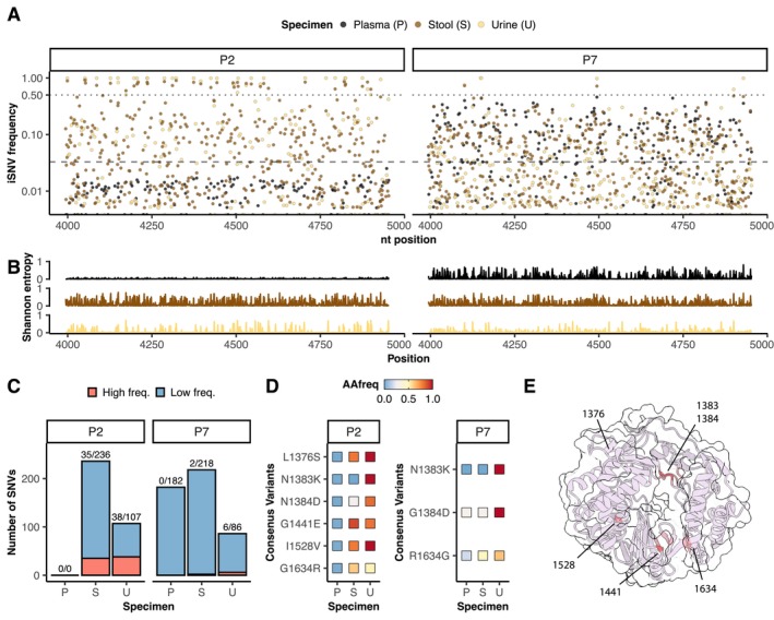
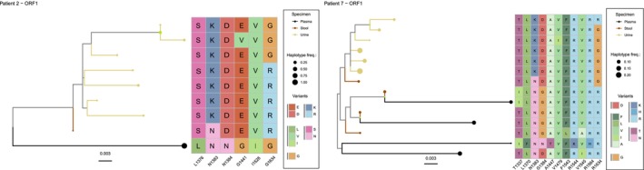
Methods: We use HEV cell culture models to study the replication cycle and the effect of antivirals in human kidney cell lines and primary cells. In addition, we identified patients with chronic HEV infection (n = 9) from which we then sequenced the viral RNA of urine, stools and plasma to analyse the viral sequence composition, to assess intra-host diversity and compartmentalisation (n = 2).

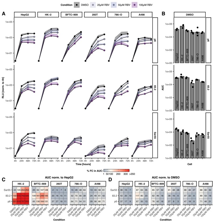
Results: A wide range of human kidney cell lines as well as primary cells supports viral entry, replication and propagation of HEV in vitro. Interestingly, the broad-spectrum antiviral ribavirin was less effective in inhibiting HEV replication in some kidney cells. Sequencing of HEV RNA-directed RNA polymerase coding region from plasma, stool and urine and subsequent phylogenetic analysis revealed diversification of HEV into tissue-specific viral subpopulations. In particular, the viruses derived from urine were found to be distinct from those derived from plasma and stool.

Conclusions: In conclusion, kidney cells support the propagation of HEV in vitro and exhibit reduced sensitivity to antiviral treatment. Furthermore, HEV patient-derived sequences demonstrated compartmentalisation into distinct clusters that correlated with sample source. Collectively, these data indicate the potential for extrahepatic replication of HEV, which may result in clinically significant disease or serve as a reservoir for patient relapse.

Trial registration: HepNet-SofE study (NCT03282474).

Keywords: compartmentalization; hepatitis E; kidney impairment.


Conflict of interest statement

The authors declare no conflicts of interest.

Figures

FIGURE 1
FIGURE 1
Recapitulation of the complete HEV replication cycle in human cells. (A) HEV replication in human renal cell lines using the Kernow‐C1/p6, 83–2‐27 and Sar55 replicon systems. Relative light units (RLU) were normalised to 4 h post‐electroporation (EPO) to quantify HEV replication. Ribavirin (RBV) was applied at concentrations of 25, 50 or 100 μM (purple shades) 4 h after electroporation. Dimethylsulfoxide (DMSO) was added at the same concentrations as a loading control (black). The lines represent data from at least three independent experiments, with the standard deviation shown as ribbon. (B) Area under the curve (AUC) quantification of replicative capacity over 72 h for DMSO control. (C) AUC normalisation of the replication signal to HepG2, allowing comparison of replication capacity between cell lines. (D) AUC normalisation to the DMSO loading control to assess cell‐type‐specific ribavirin inhibition.
FIGURE 2
FIGURE 2
HEV can complete its full viral replication cycle in kidney cells. (A) Viral antigen production was measured after electroporation of viral RNA into kidney and liver cells using the Wantai antigen enzyme‐linked immunosorbent assay (ELISA). (B) Biophysical properties of the virus produced in different renal cell lines were assessed by measuring the RNA content after density centrifugation of cell lysates and supernatants. (C) Infectivity of virus particles produced in renal cell lines was assessed in HepG2/C3A cells by determining focus‐forming units (FFU) per mL of virus stock. (D) Susceptibility of renal cells to HEV infection was tested using neHEV particles produced in HepG2 cells. Five days after infection, the cells were immunostained for the ORF2‐encoded capsid protein (CP) using an anti‐capsid protein antibody. An anti‐HEV antibody (ab) at a concentration of 10 μg/mL was used as a specificity control. (E) Ability of neHEV produced in kidney cells to infect the same cell type was assessed.
FIGURE 3
FIGURE 3
Virus RNA sequencing shows divergence of HEV populations in samples from different sources. (A) Intra‐host single nucleotide frequencies (iSNV) of the RNA‐directed RNA polymerase (RdRp) coding region of virus populations sampled from plasma (P, black), stool (S, brown) and urine (U, yellow) of patient 2 (P2) and patient 7 (P7). (B) Nucleotide sequence divergence summarised as Shannon entropy. (C) Quantification of low frequency (above 3.28%, blue) and high frequency (above 50%, red). (D) Frequency of amino acid (AA) substitutions within the RdRp of HEV strains in the three samples of both patients. (E) Representation of amino acid substitutions on an alpha‐fold‐2‐generated 3D structure model of the viral polymerase.
FIGURE 4
FIGURE 4
Phylogenetic reconstruction from haplotype data. NGS reads were used to reconstruct haplotypes using CliqueSNV. A multiple sequence alignment was constructed using Clutal Omega. Phylogenetic trees were reconstructed using the maximum likelihood method implemented in IQ‐tree 2. The trees were then visualised in R using the ggtree package. Supported bootstrap values (≥ 80%) are visualised with green dots on the parent branches. Finally, heatmaps were created to visualise amino acid substitutions in comparison to the reference sequence. The colour coding of the heatmap depicts chemical properties, with negative charges (D, E), positive charges (R, H, K), polar‐uncharged (S, T, N), non‐polar‐uncharged (A, F, I, L, V) and others (C, P, G) represented by different colours. The colour of the dots indicates the specimen origin, with yellow dots representing urine, brown dots representing stool and black dots representing plasma. The size of the dots indicates the frequency of haplotypes.

Similar articles

References

    1. World Health Organization , “Hepatitis E Fact Sheets [Internet],” (2022) cited 2023 Jan 3, https://www.who.int/news‐room/fact‐sheets/detail/hepatitis‐e.
    1. Nimgaonkar I., Ding Q., Schwartz R. E., and Ploss A., “Hepatitis E Virus: Advances and Challenges,” Nature Reviews. Gastroenterology & Hepatology 15, no. 2 (2018): 96–110. - PMC - PubMed
    1. Dalton H. R., Kamar N., Baylis S. A., Moradpour D., Wedemeyer H., and Negro F., “EASL Clinical Practice Guidelines on Hepatitis E Virus Infection,” Journal of Hepatology 68, no. 6 (2018): 1256–1271. - PubMed
    1. van Tong H., Hoan N. X., Wang B., Wedemeyer H., Bock C. T., and Velavan T. P., “Hepatitis E Virus Mutations: Functional and Clinical Relevance,” eBioMedicine 1, no. 11 (2016): 31–42. - PMC - PubMed
    1. Gömer A., Klöhn M., Jagst M., et al., “Emergence of Resistance‐Associated Variants During Sofosbuvir Treatment in Chronically Infected Hepatitis E Patients,” Hepatology 78, no. 6 (2023): 1882–1895. - PMC - PubMed

Publication types

Associated data

LinkOut - more resources