. 2025 Sep;27(9):101506.
doi: 10.1016/j.gim.2025.101506. Epub 2025 Jun 23.

ELFN1 deficiency: The mechanistic basis and phenotypic spectrum of a neurodevelopmental disorder with epilepsy

Collaborators, Affiliations

ELFN1 deficiency: The mechanistic basis and phenotypic spectrum of a neurodevelopmental disorder with epilepsy

Rhys Dore et al. Genet Med. 2025 Sep.

Abstract

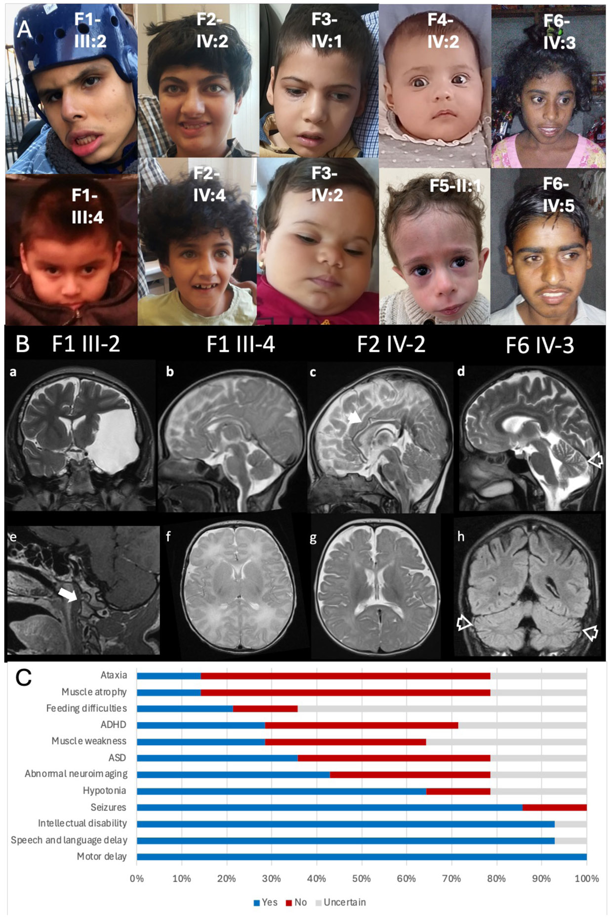
Purpose: Synaptic communication deficits are central to many neurodevelopmental disorders. However, for rare monogenic conditions, these disorders remain poorly defined, with limited understanding of their molecular etiology. A homozygous frameshift variant in the synaptic cell adhesion molecule ELFN1 was reported in a family with 3 affected siblings with epileptic encephalopathy, alongside a missense variant of uncertain significance in a cohort study involving a family with intellectual disability. Therefore, we sought to evaluate the role and mechanism of biallelic ELFN1 variants in disease pathogenesis.

Methods: We describe 8 newly identified individuals from 5 unrelated families, all carrying homozygous ELFN1 variants, including frameshift and in-frame deletions. By integrating data from these cases with clinical details from 6 previously reported individuals, we delineate the phenotypic spectrum associated with ELFN1 variants.

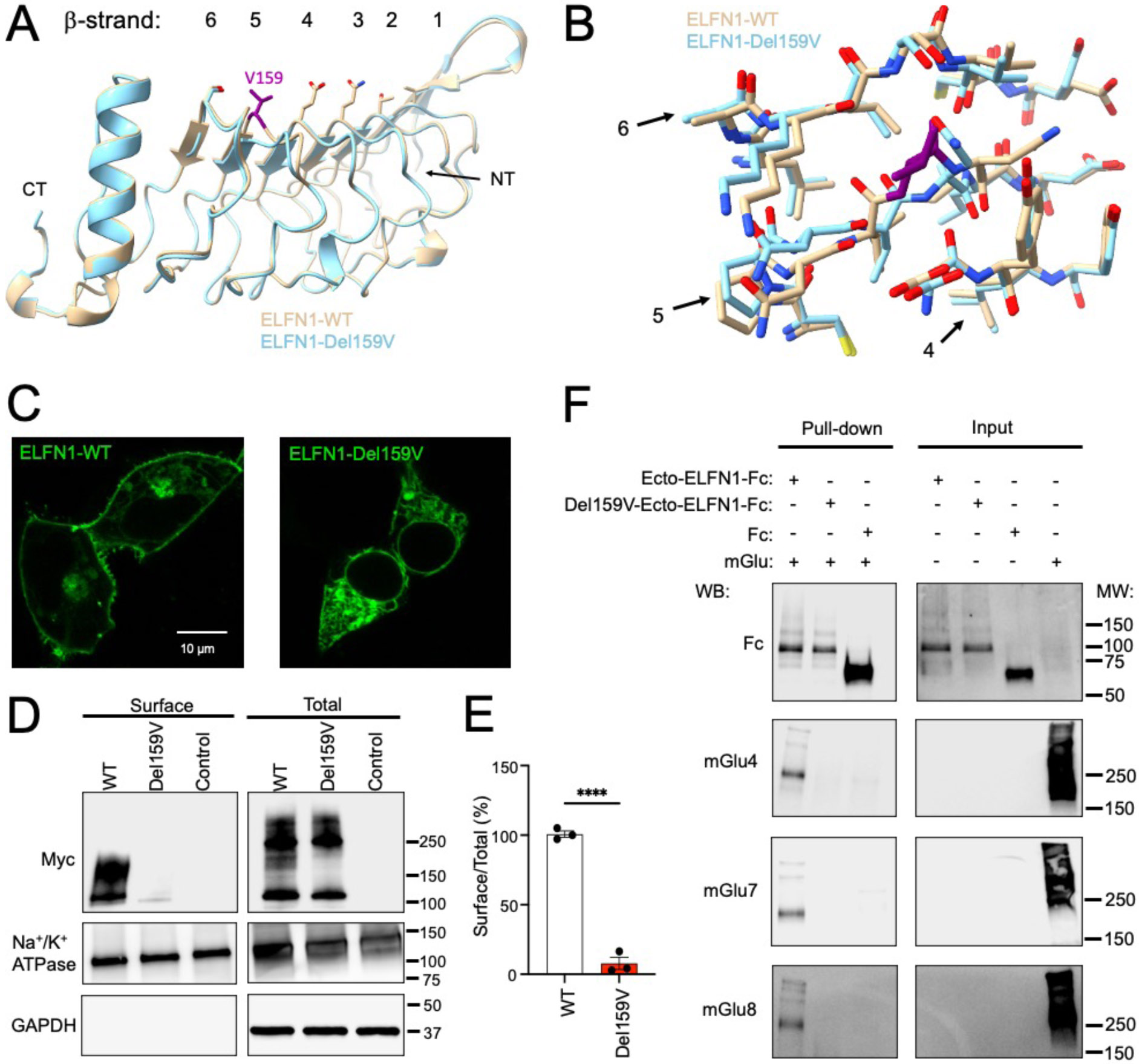
Results: Clinical features include varying degrees of developmental delay/intellectual disability, epilepsy, and movement disorders. Molecular investigations reveal that these variants disrupt ELFN1 protein trafficking to the cell surface, resulting in loss of function. Functional modeling in mice and zebrafish demonstrates the role of Elfn1 loss in motor activity abnormalities and seizures.

Conclusion: Our findings establish ELFN1 deficiency as the cause of a distinct, rare neurodevelopmental disorder, providing a foundation for future investigations into its pathophysiology and therapeutic strategies.

Keywords: ELFN1; Epilepsy; Movement disorder; Neurodevelopmental disorder; Synaptic adhesion protein.


Conflict of interest statement

Conflict of Interest The authors declare no conflicts of interest.

Figures

Figure 1.
Figure 1.. Pedigrees and variant visualization
A) Pedigrees demonstrating the seven families and genotypes of tested individuals, indicated using + (pathogenic variant) and – (wild type). B) The ELFN1 transcript (NM_001128636.4) with each exon numbered 1-4 and the coding region depicted in dark blue. The pathogenic deletions identified in this report are represented at their location along the transcript in black text, whilst the previously identified pathogenic deletion is presented in grey text. C) The ELFN1 protein with functional regions depicted by colour and labelled as SP (signal peptide), LRR (leucine rich repeat), LRRCT (leucine rich C-terminal domain), FN3 (fibronectin type III domain), TM (transmembrane domain). The pathogenic deletions identified in this report are represented at their location along the transcript in black text, whilst the previously identified pathogenic deletion is presented in grey text. Regions of putatively translated frameshifts are demonstrated by an extended red line, whilst the downstream new stop codon is shown via a red cross.
Figure 2.
Figure 2.. Facial photographs and neuroimaging of patients.
A) Facial photographs of the reported cases. B) Brain MRI. Family 1 III- 2 Coronal T2 weighted image (WI) and Sagittal post gad T1WI (a and e) shows arachnoid cyst in the left middle cranial fossa with mass effect (a) and atlantooccipital subluxation (arrow, e). Family 1 III-4 Sagittal and axial T2WI (b and e) showing normal morphology and signal changes of the brain. Family 2 IV-2 Sagittal and axial T2WI (c and g) showing mild diffuse volume loss with slightly thin corpus callosum (arrowhead, c). Family 6 IV-3 Sagittal T2WI and coronal FLAIR (d and h) showing brain volume loss including involvement of the cerebellar vermis and cerebellar hemispheres (open arrowheads, d and h). C) Common clinical features exhibited by patients, demonstrated by percentage affected.
Figure 3.
Figure 3.. Molecular mechanisms of ELFN1 disruption caused by Pro50His, Val591fs, and Gln508fs variants.
A) Synaptic localization of Elfn1. B) Representative Western Blots of surface and total protein expression level of c.149C>A p.(Pro50His) (P50H), c.1767del p.(Val591CysfsTer93) (Fs591), and c.1522del p.(Gln508SerfsTer176) (Fs508) ELFN1. C) Quantification of the ratio of surface to total protein expression level. D) Representative confocal images showing the WT, Pro50His, Val591fs, and Gln508fs ELFN1 expressed in HEK293 cells.
Figure 4.
Figure 4.. Structural model and functional effects of valine ELFN1 Val159del
A) Overlay of LRR region of AlphaFold generated structures of WT ELFN1 and ELFN1 c.475_477del p.(Val159del) (Del159V). Position of valine 159 is shown by purple stick model. B) Detailed analysis of backbone and side chain configurations in β-strands of LRR domain in WT ELFN1 and ELFN1 Val159del. C) Analysis of WT and c.475_477del p.(Val159del) ELFN1 Venus localization by confocal microscopy in live HEK293 cells. Representative confocal images are shown. D) Examination of ELFN1 expression and surface localization by biotinylation. Representative Western blots of total and surface content of WT and c.475_477del p.(Val159del) ELFN1 are shown. E) Quantification of the surface biotinylation experiments shown in panel B from three independent experiments. ****p<0.0001, t-test. F) Representative western blots with inputs and pull-downs captured by Ecto-ELFN1-Fc and Val159del-Ecto-ELFN1-Fc by protein G beads.
Figure 5.
Figure 5.. Studying ELFN1 deficiency in animal models.
A) Time-course of locomotor activity of wildtype, Elfn1+/−, knockout Elfn1−/− and heterozygous Elfn+/− mice in the open field. B) Quantification of the total locomotor activity in the open filed over 2 hours. C) Behavioral and (D,E,F) electrophysiological seizure analysis of MO-injected zebrafish larvae at 5 dpf. C) Behavioral data is compared to control MO (elfn1a+/+/elfn1b+/+) and expressed as cumulative duration in highly active state (mean ± SEM) (n control MO = 73, n elfn1a/elfn1b MO = 29-30). D,E,F) Electrophysiological epileptiform activity is assessed via non-invasive local field potential recordings (n control MO = 18, n elfn1a/elfn1b MO = 15-16). D) Data is visually analysed and expressed as the number of epileptiform events (mean ± SEM) and compared to control MO. E,F) Data is normalized against control MO and expressed as (E) normalized power spectral density (PSD) (mean ±SEM) per larva per 10 Hz frequency band from 1-150 Hz and as (F) normalized PSD (mean ±SEM) per larva within the 10-90 Hz region. Statistical analysis was performed using (C,D,E,F) non-parametric Kruskal-Wallis test with Dunn’s multiple comparisons test. Outliers were removed via the ROUT test (Q = 1%). Significance levels: * p ≤ 0.05; ** p ≤ 0.01; *** p ≤ 0.001; **** p ≤ 0.0001.

References

    1. Südhof TC. The cell biology of synapse formation. J Cell Biol. 2021;220(7). 10.1083/jcb.202103052 - DOI - PMC - PubMed
    1. Kurshan PT, Shen K. Synaptogenic pathways. Curr Opin Neurobiol. 2019;57:156–162. 10.1016/j.conb.2019.03.005 - DOI - PubMed
    1. Sheng M, Hoogenraad CC. The postsynaptic architecture of excitatory synapses: a more quantitative view. Annu Rev Biochem. 2007;76:823–847. 10.1146/annurev.biochem.76.060805.160029 - DOI - PubMed
    1. Magee JC, Grienberger C. Synaptic plasticity forms and functions. Annu Rev Neurosci. 2020;43:95–117. 10.1146/annurev-neuro-090919-022842 - DOI - PubMed
    1. Turrigiano G. Homeostatic synaptic plasticity: local and global mechanisms for stabilizing neuronal function. Cold Spring Harb Perspect Biol. 2012;4(1):a005736. 10.1101/cshperspect.a005736 - DOI - PMC - PubMed

Substances