. 2025 Dec;17(1):2519706.
doi: 10.1080/19490976.2025.2519706. Epub 2025 Jun 27.

Gut microbial metabolite butyrate suppresses hepatocellular carcinoma growth via CXCL11-dependent enhancement of natural killer cell infiltration

Affiliations

Gut microbial metabolite butyrate suppresses hepatocellular carcinoma growth via CXCL11-dependent enhancement of natural killer cell infiltration

Menghan Zhang et al. Gut Microbes. 2025 Dec.

Abstract

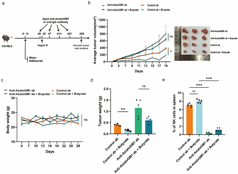
Gut microbiota-derived butyrate plays a vital role in attenuating hepatocellular carcinoma (HCC) in murine models. However, the precise molecular mechanisms by which butyrate exerts its effects are largely undefined. Plasma short-chain fatty acids (SCFAs) were quantitatively measured by using gas chromatography-mass spectrometry (GC-MS) to access their association with HCC prognosis. Tumor-infiltrating immune cells were characterized by flow cytometry. The interactions between butyrate and natural killer (NK) cells were studied using in vitro assays, including migration, cytotoxic degranulation, and co-culture experiments. In vivo validation was conducted through neutralization experiments. The molecular pathways regulated by butyrate were further investigated by employing RNA sequencing (RNA-seq), chromatin immunoprecipitation sequencing (ChIP-seq), and Assay for Transposase-Accessible Chromatin using sequencing (ATAC-seq). A positive correlation was observed between elevated plasma butyrate levels and improved prognosis in HCC patients. Notably, butyrate inhibited tumor progression by enhancing NK cell infiltration into tumor tissues. Mechanistically, butyrate stimulated cytokine secretion, notably significantly enhancing the production of the chemokine CXCL11, thereby facilitating NK cell infiltration. Gene Set Enrichment Analysis (GSEA) of hepatic tumor cell lines revealed that the chemokine signaling and NK cell-mediated cytotoxicity pathways were upregulated following butyrate stimulation. Furthermore, transcriptomic and epigenomic analyses showed that exposure to butyrate induced de novo chromatin accessibility and enhancer remodeling, regulated by STAT family transcription factors. Our study demonstrated that butyrate was able to enhance the expression of CXCL11. This is likely attributed to chromatin remodeling, and then promoting NK cell infiltration and exerting effective anti-tumor effects on HCC.

Keywords: Gut microbiota; butyrate; epigenetic; hepatocellular carcinoma; natural killer cells.


Conflict of interest statement

No potential conflict of interest was reported by the author(s).

Figures

None
Graphical abstract
Figure 1.
Figure 1.
Gut microbiota-derived butyrate is associated with the prognosis of HCC patients. (a) Heatmap illustrating alterations in human plasma short-chain fatty acids (SCFAs) in healthy donors (HD, N = 20) and hepatocellular carcinoma patients (HCC, N = 36). (b) Spearman correlation analysis showing the association between the concentration of plasma SCFAs and clinical characteristics in HCC patients. *p < 0.05, **p < 0.01, and ***p < 0.001, by Spearman test. (c) Progression-free survival in butyrate-high and butyrate-low groups. p<0.001, by log-rank test. (d) GC-MS analysis revealing differences in plasma butyrate level between HD (N = 20) and HCC (N = 36) groups. (e) Receiver operating characteristic (ROC) analysis distinguishing HCC patients and HD in terms of plasma butyrate. (f) Percentage of community abundance at the phylum and genus level of gut microbiota in the two studied groups. **p < 0.01, by Mann-Whitney test.
Figure 2.
Figure 2.
Butyrate exposure significantly attenuates tumor growth and reshapes the tumor microenvironment. (a) Schematic overview of butyrate treatment in Hepa1–6-bearing mice. N = 5 mice for each group. (b) Tumor volume was monitored, and representative images shown on the right. N = 5 mice per group. (c) Representative images of Ki67-positive cells of tumor sections in the control and butyrate-treated group (scale bar, 100 μM). Quantification of Ki67+ in the tumor (right). N = 5 mice per group. (d) C57BL/6 mice were pretreated with or without butyrate feeding as described in methods. Tumor-infiltrating immune cells were measured by flow cytometry. N = 5 mice per group. Data are expressed as mean ± SEM. *p < 0.05, **p < 0.01, by two-tailed Student’s t-test. (e) Immunofluorescence staining of tumor tissues with anti- NCR1.
Figure 3.
Figure 3.
NK cells are essential for butyrate-mediated suppression of hepatic tumor growth in a mouse model. (a) Schematic illustration of the experimental design and timeline for NK cell depletion assays in mouse models. (b) Monitoring of average tumor sizes under different conditions since tumor inoculation. N = 5 mice for each group. Representative images are displayed on the right. (c) Weight curves for mice. N = 5 mice for each group. (d) Recording of average tumor weight. N = 5 mice for each group. (e) Flow cytometrical measurement of the relative percentage (%) of NK cells in spleens from mice. N = 5 mice for each group. Data are presented as mean ± SEM. *p < 0.05, **p < 0.01, ***p < 0.001, two-tailed Student’s test.
Figure 4.
Figure 4.
Butyrate-mediated up-regulation of CXCL11 is crucial for NK cell migration. (a) Schematic illustration of NK cell migration assays. (b) Transwell assay showing the NK cells migrating towards conditioned media from HepG2 and Huh7 cells pre-treated with butyrate (1 mm) and PBS for 48 h. (c) RNA-seq analysis of differentially-expressed genes in butyrate-treated hepatic tumor cells (HepG2) for 48 h, with a heatmap illustrating significantly upregulated genes. (d) Gene set enrichment analysis (GSEA) based on RNA-seq profiles, showing a positive association between butyrate treatment and the chemokines signaling pathway and the natural killer cells-mediated cytotoxicity pathway. (e) Analysis of conditioned media from HepG2 cells treated with butyrate or PBS using a 92 inflammation panels, with quantitative assessment of CXCL11 expression displayed using normalization protein expression (NPX) data. (f) qRT-PCR measurement of relative CXCL11 mRNA expression in HepG2 and Huh7 cells treated with butyrate (1 mm) for 48 h compared to PBS-treated cells, with ACTINB as the internal reference. (g) ELISA measurement of CXCL11 protein levels in supernatants from HepG2 and Huh7 cells treated with butyrate (1 mm) or PBS for 48 h. (h) Average number of NK cells that migrated toward conditioned media with or without recombinant human CXCL11 protein. (i) Average number of NK cells that migrated toward conditioned media collected from HepG2 and Huh7 cells that pretreated with butyrate, with or without CXCL11-neutralizing antibodies. Data are given as mean ± SEM based on three independent experiments. *p < 0.05; **p < 0.01; ***p < 0.001, two-tailed Student’s test.
Figure 5.
Figure 5.
Histone modifications at gene enhancer sites modulate CXCL11 expression. (a) HepG2G2 cells were stimulated with or without butyrate for 48 h and analyzed for changes in ATAC-seq. Signal coverage heatmaps of ATAC-seq results are shown. (b) Principal component analysis (PCA) of clusters according to (a). (c) Genome browser view of normalized signals of H3K9ac, H3K27ac ChIP-seq and ATAC-seq at the CXCL11 locus in HepG2 cells with or without butyrate treatment. (d) Heatmap showing relative frequency of TF consensus motifs among five different classes defined in (a). Main TF families are highlighted at the top. Motif analysis was conducted using HOMER and visualized with R. (e) Genome browser view of normalized signals of H3K9ac, H3K27ac ChIP-seq and ATAC-seq at the locus of CXCL11 in HepG2 cells with or without butyrate treatment. (f – g) ChIP-qPCR performed in HepG2 and Huh7 cells treated with butyrate to detect the enrichment levels of CXCL11 enhancer using H3K27ac and H3K9ac antibodies and IgG control. (h) The CXCL11 enhancer region is highly enriched with H3K27ac. The sites of the putative STAT4 binding motif in the − 76053933 CXCL11 enhancer were predicted using the JASPAR database. (i) Gel electrophoresis of STAT4 enrichment at binding sites in the CXCL11 enhancer. (j) Luciferase assay of HEK293 T cells transfected for 48 h with the indicated plasmids: PGL3-basic, CXCL11 promoter and CXCL11 promoter + enhancer plasmid. Data are presented as mean ± SEM based on three independent experiments. *p < 0.05; **p < 0.01; ***p < 0.001, by two-tailed Student’s t-test.
Figure 6.
Figure 6.
Butyrate-mediated re-expression of CXCL11 is necessary for tumor suppression in hepatic tumor mouse model. (a) Schematic representation of the experimental design for CXCL11-neutralizing assays and timeline of mouse models. (b) Average tumor sizes were monitored for the indicated conditions since tumor inoculation. N = 5 mice for each group. Representative images are shown at right. (c) Mice weight curves are monitored. N = 5 mice for each group. (d) Average tumor weight was recorded. N = 5 mice for each group. (e) The percentage of Hepa1–6 tumor-infiltrating NK cell was flow cytometrically measured. N = 5 mice for each group. Data are presented as the mean ± SEM; ns, not significant; *p < 0.05, **p < 0.01, ***p < 0.001, and ****p < 0.0001, two-tailed Student’s test.

References

    1. Waked I, Alsammany S, Tirmazy SH, Rasul K, Bani-Issa J, Abdel-Razek W, Omar A, Shafik A, Eid S, Abdelaal A, et al. Multidisciplinary consensus recommendations for management of hepatocellular carcinoma in Middle East and North Africa region. Liver Int. 2023;43(10):2062–16. doi: 10.1111/liv.15685. - DOI - PubMed
    1. Gao S, Jiang X, Wang L, Jiang S, Luo H, Chen Y, Peng C.. The pathogenesis of liver cancer and the therapeutic potential of bioactive substances. Front Pharmacol. 2022;13:1029601. doi: 10.3389/fphar.2022.1029601. - DOI - PMC - PubMed
    1. Ringelhan M, Pfister D, O’Connor T, Pikarsky E, Heikenwalder M. The immunology of hepatocellular carcinoma. Nat Immunol. 2018;19(3):222–232. doi: 10.1038/s41590-018-0044-z. - DOI - PubMed
    1. Song Q, Zhang X, Liu W, Wei H, Liang W, Zhou Y, Ding Y, Ji F, ho-Kwan Cheung A, Wong N, et al. Bifidobacterium pseudolongum-generated acetate suppresses non-alcoholic fatty liver disease-associated hepatocellular carcinoma. J Hepatol. 2023;79(6):1352–1365. doi: 10.1016/j.jhep.2023.07.005. - DOI - PubMed
    1. Gopalakrishnan V, Spencer CN, Nezi L, Reuben A, Andrews MC, Karpinets TV, Prieto PA, Vicente D, Hoffman K, Wei SC, et al. Gut microbiome modulates response to anti-PD-1 immunotherapy in melanoma patients. Science. 2018;359(6371):97–103. doi: 10.1126/science.aan4236. - DOI - PMC - PubMed

MeSH terms

LinkOut - more resources