The Sialome of the Retina, Alteration in Age-Related Macular Degeneration Pathology, and Potential Impacts on Complement Factor H
- PMID: 40576434
- PMCID: PMC12212450
- DOI: 10.1167/iovs.66.6.81
The Sialome of the Retina, Alteration in Age-Related Macular Degeneration Pathology, and Potential Impacts on Complement Factor H
Abstract
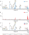
Purpose: Little is known about sialic acids of the human retina, despite their integral role in self /non-self-discrimination by complement factor H (FH), the alternative complement pathway inhibitor.
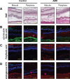
Methods: A custom sialoglycan microarray was used to characterize the sialic acid-binding specificity of native FH or recombinant molecules where IgG Fc was fused to FH domains 16 to 20 (which contains a sialic acid-binding site), domains 6 and 7 (which contains a glycosaminoglycan-binding site), or the FH-related proteins (FHRs) 1 and 3. We analyzed macular and peripheral retinal tissue from postmortem ocular globes for the amount, type, and presentation (glycosidic linkage type) of sialic acid in individuals with age-related macular degeneration (AMD) and age-matched controls using fluorescent lectins and antibodies to detect sialic acid and endogenous FH. Released sialic acids from neural retina, retinal pigmented epithelium (RPE) cells, and the Bruch's membrane (BrM) were labeled with 1,2-diamino-4,5-methylenedioxybenzene-2HCl (DMB), separated and quantified by high-performance liquid chromatography (HPLC).
Results: Both native FH and the recombinant FH domains 16 to 20 recognized Neu5Ac and Neu5Gc that is α2-3-linked to the underlying galactose. 4-O-Actylation of sialic acid and sulfation of GlcNAc did not inhibit binding. Different linkage types of sialic acid were localized at different layers of the retina. The greatest density of α2-3-sialic acid, which is the preferred ligand of FH, did not colocalize with endogenous FH. The level of sialic acids at the BrM/choroid interface of the macula and peripheral retina of individuals with AMD were significantly reduced.
Conclusions: The sialome of the human retina is altered in AMD. This may affect FH binding and, consequently, alternative complement pathway regulation.
Conflict of interest statement
Disclosure:
Figures


Update of
-
The sialome of the retina, alteration in age-related macular degeneration (AMD) pathology and potential impacts on Complement Factor H.bioRxiv [Preprint]. 2025 Mar 13:2025.03.09.642149. doi: 10.1101/2025.03.09.642149. bioRxiv. 2025. Update in: Invest Ophthalmol Vis Sci. 2025 Jun 2;66(6):81. doi: 10.1167/iovs.66.6.81. PMID: 40161805 Free PMC article. Updated. Preprint.
References
MeSH terms
Substances
Grants and funding
LinkOut - more resources
Full Text Sources
Medical
Miscellaneous

