

Association of Pelvic Structure Involvement and Tumor Morphology at MRI with Prognosis Following Resection in Locally Recurrent Rectal Cancer
- PMID: 40576488
- PMCID: PMC12304544
- DOI: 10.1148/rycan.240246
Association of Pelvic Structure Involvement and Tumor Morphology at MRI with Prognosis Following Resection in Locally Recurrent Rectal Cancer
Abstract
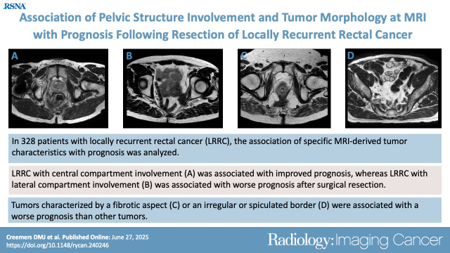
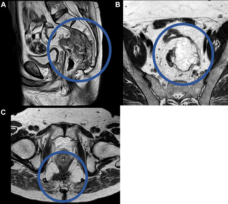
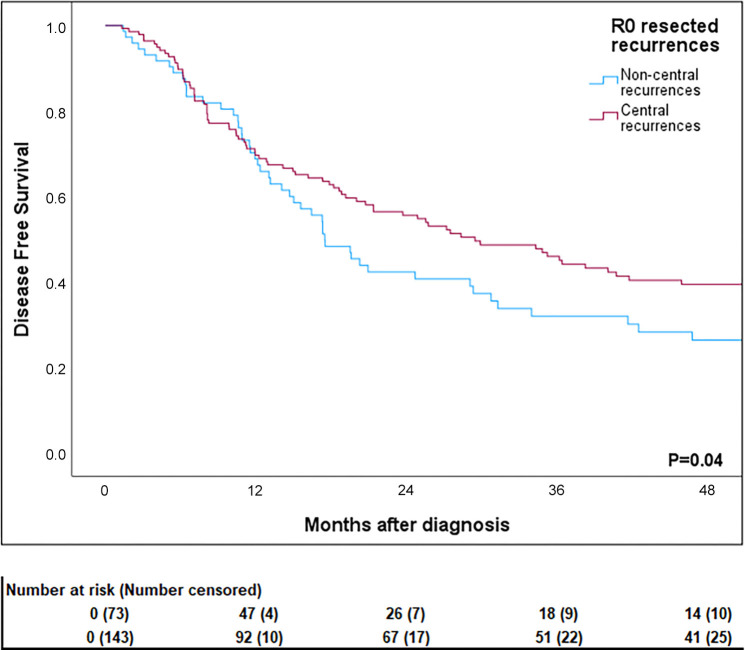
Purpose To determine the influence of location, extent of tissue invasion, and tumor morphology at MRI on the resectability of locally recurrent rectal cancer (LRRC) and postresection oncologic outcomes of LRRC. Materials and Methods This retrospective observational study included consecutive patients diagnosed with LRRC who underwent surgery with curative intent at the Catharina Hospital Eindhoven and Karolinska University Hospital Stockholm between January 2003 and December 2017. Two expert radiologists reviewed available MR images while adhering to a standardized reviewing checklist. The effect of pelvic structure involvement, tumor morphology on the primary outcome of resection margin status, and secondary outcomes of overall survival and disease-free survival were assessed using univariable and multivariable logistic regression and Cox proportional hazard analyses. Results The final analysis included 328 patients with LRRC (mean age ± SD, 64.9 years ± 9.6; 126 female, 202 male). Resection margins were negative in 217 (66.2%) patients and positive in 111 patients (33.8%). Tumor size, tumor type, and border type on MR images were all associated with resectability. Central recurrences were associated with the lowest likelihood of positive resection margins (odds ratio [OR], 0.45; 95% CI: 0.28, 0.71; P < .001), whereas lateral recurrences were associated with the highest likelihood (OR, 2.00; 95% CI: 1.25, 3.19: P = .004). Similarly, central recurrences were associated with better disease-free survival compared with lateral recurrences (hazard ratio [HR], 0.69; 95% CI: 0.53, 0.90; P = .006 vs HR, 1.49; 95% CI: 1.14, 1.94; P = .003, respectively). Similar findings were observed after correcting for resection margin status. Conclusion Standardized MRI assessment of tumor characteristics in patients with LRRC resulted in the identification of specific prognostic factors. Central compartment involvement and well-defined tumors were associated with improved prognosis, whereas lateral compartment involvement and fibrotic spiculated tumors were associated with a worse prognosis after surgical resection. Keywords: Rectum, MR-Imaging, Abdomen/GI, Oncology, Surgery, Locally Recurrent Rectal Cancer, Tumor Biology Supplemental material is available for this article. © RSNA, 2025.
Keywords: Abdomen/GI; Locally Recurrent Rectal Cancer; MR-Imaging; Oncology; Rectum; Surgery; Tumor Biology.
Conflict of interest statement
Figures


Similar articles
-
Is the Thickness of the Margin Associated With Local Recurrence and Survival in Patients With Myxofibrosarcoma?Clin Orthop Relat Res. 2023 Nov 1;481(11):2125-2136. doi: 10.1097/CORR.0000000000002709. Epub 2023 May 29. Clin Orthop Relat Res. 2023. PMID: 37249339 Free PMC article.
-
Oncological Outcomes of Intersphincteric Resection Versus Abdominoperineal Resection for ypT3 Low Rectal Cancer After Neoadjuvant Chemoradiotherapy: A Multicenter Retrospective Analysis.Dis Colon Rectum. 2025 Aug 1;68(8):951-961. doi: 10.1097/DCR.0000000000003821. Epub 2025 May 7. Dis Colon Rectum. 2025. PMID: 40331664
-
Early failure following pelvic exenteration: Who are the bad actors?Surgeon. 2025 Aug;23(4):211-215. doi: 10.1016/j.surge.2025.02.010. Epub 2025 Mar 14. Surgeon. 2025. PMID: 40087056
-
A systematic review of the pathological determinants of outcome following resection by pelvic exenteration of locally advanced and locally recurrent rectal cancer.Int J Surg. 2022 Aug;104:106738. doi: 10.1016/j.ijsu.2022.106738. Epub 2022 Jul 1. Int J Surg. 2022. PMID: 35781038
-
A Systematic Review to Assess Resection Margin Status After Abdominoperineal Excision and Pelvic Exenteration for Rectal Cancer.Ann Surg. 2017 Feb;265(2):291-299. doi: 10.1097/SLA.0000000000001963. Ann Surg. 2017. PMID: 27537531
References
-
- Westberg K , Palmer G , Hjern F , Johansson H , Holm T , Martling A . Management and prognosis of locally recurrent rectal cancer – A national population-based study . Eur J Surg Oncol 2018. ; 44 ( 1 ): 100 – 107 . - PubMed
-
- Harris CA , Solomon MJ , Heriot AG , et al . The Outcomes and Patterns of Treatment Failure after Surgery for Locally Recurrent Rectal Cancer . Ann Surg 2016. ; 264 ( 2 ): 323 – 329 . - PubMed
-
- Madoff RD . Extended resections for advanced rectal cancer . Br J Surg 2006. ; 93 ( 11 ): 1311 – 1312 . - PubMed
-
- Milne T , Solomon MJ , Lee P , Young JM , Stalley P , Harrison JD . Assessing the impact of a sacral resection on morbidity and survival after extended radical surgery for locally recurrent rectal cancer . Ann Surg 2013. ; 258 ( 6 ): 1007 – 1013 . - PubMed
-
- Austin KKS , Young JM , Solomon MJ . Quality of life of survivors after pelvic exenteration for rectal cancer . Dis Colon Rectum 2010. ; 53 ( 8 ): 1121 – 1126 . - PubMed
Publication types
MeSH terms
LinkOut - more resources
Full Text Sources
Medical
