Review
. 2025 Aug 1;329(2):H340-H357.
doi: 10.1152/ajpheart.00352.2025. Epub 2025 Jun 27.

Endothelial metabolic zonation in the vascular network: a spatiotemporal blueprint for angiogenesis

Affiliations
Review

Endothelial metabolic zonation in the vascular network: a spatiotemporal blueprint for angiogenesis

Kesha K Dalal et al. Am J Physiol Heart Circ Physiol. .

Abstract

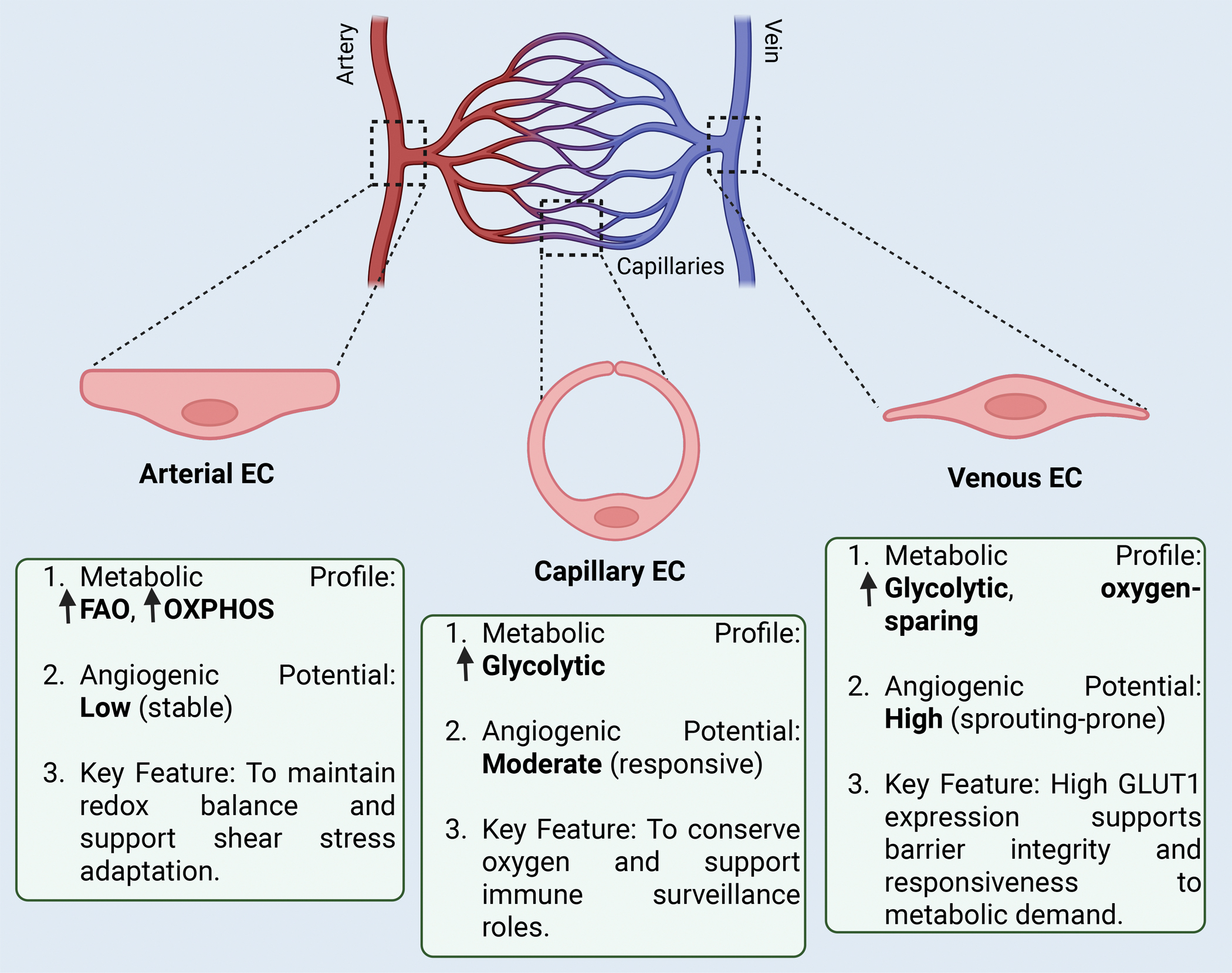
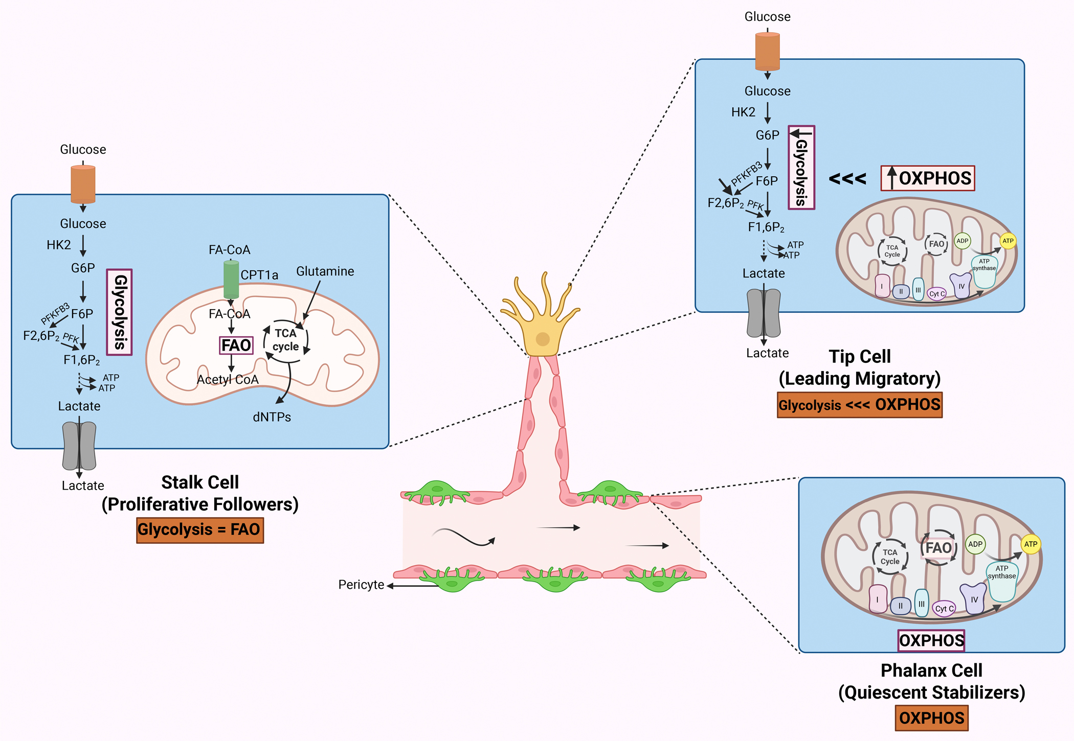
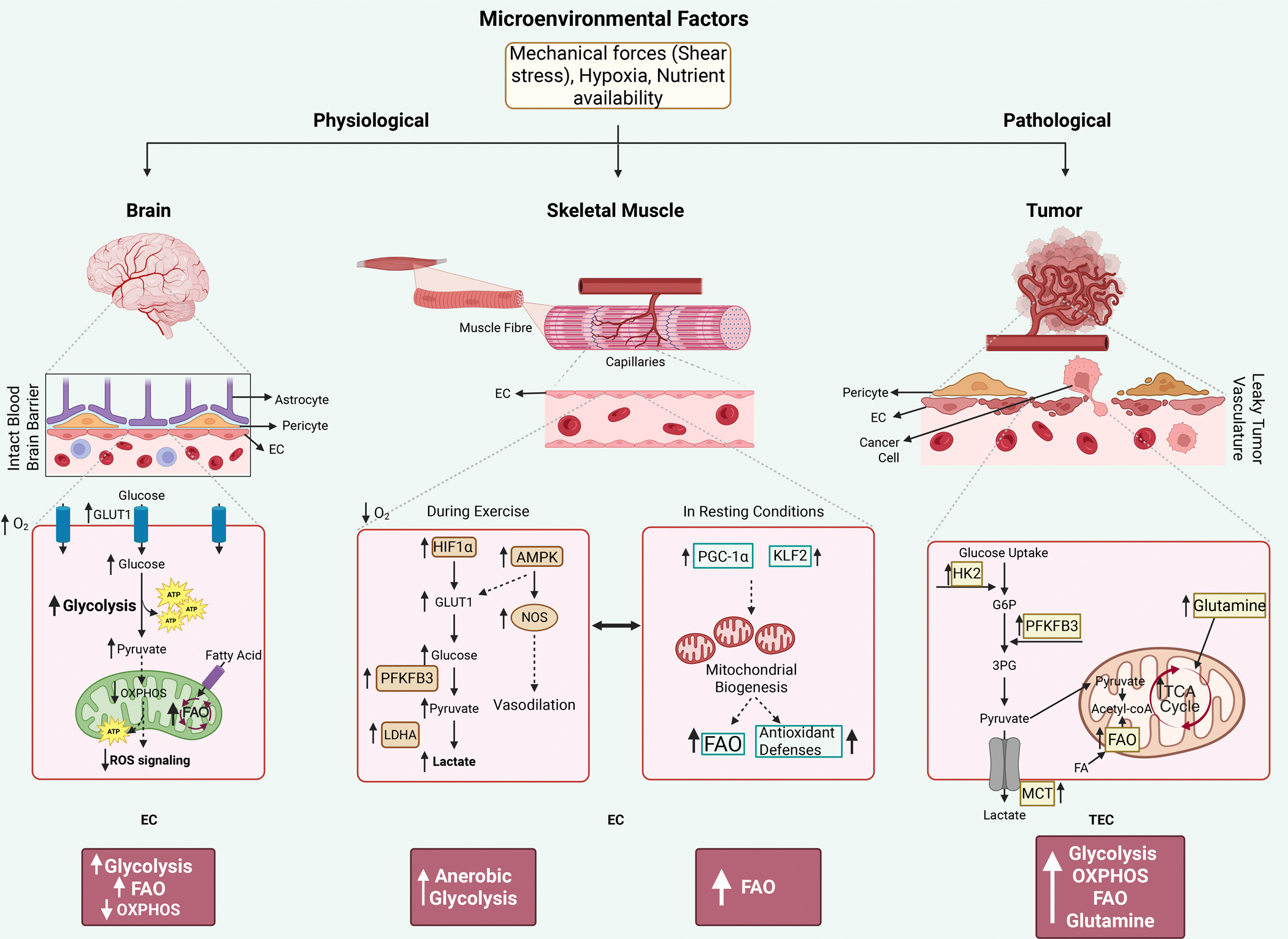
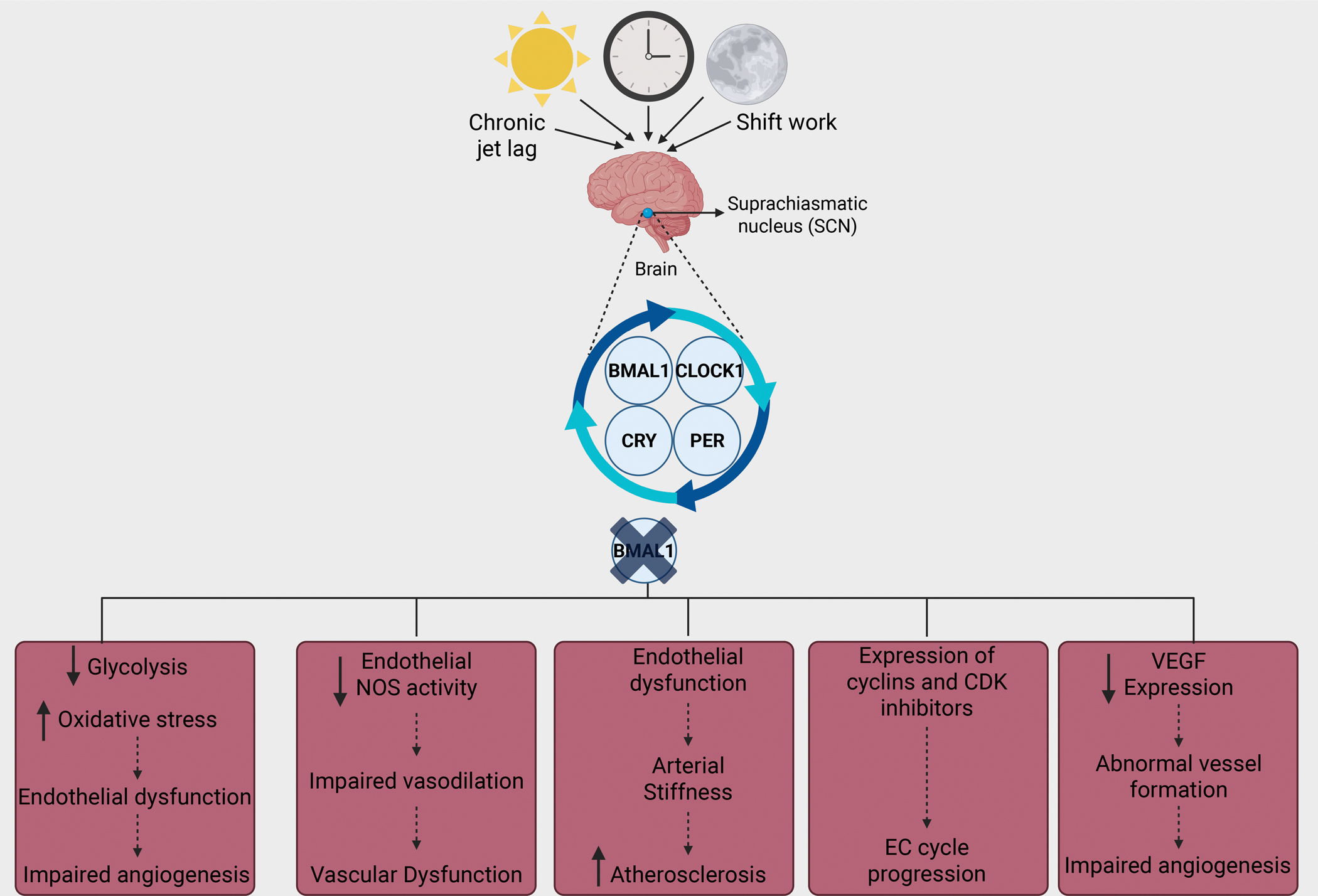
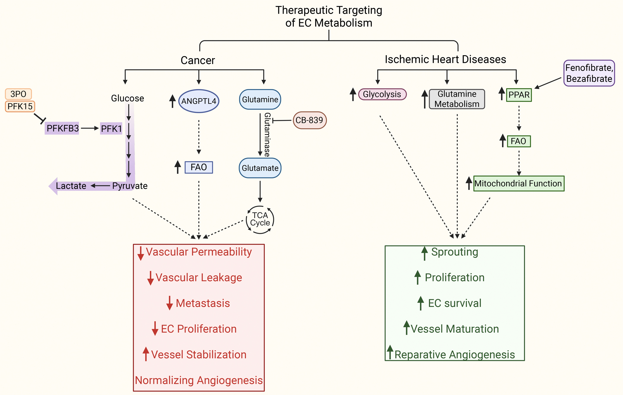
Angiogenesis, a cornerstone of vascular development, tissue regeneration, and tumor progression, is critically orchestrated by the metabolic behavior of endothelial cells (EC). Recent discoveries have redefined EC not as metabolically uniform entities, but as spatially and functionally heterogeneous populations whose metabolic states govern their angiogenic potential. This review presents a comprehensive synthesis of metabolic zonation in EC, spanning arterial, venous, and capillary domains, and highlights cell-type-specific programs during sprouting angiogenesis-including tip, stalk, and phalanx cells. We explore how distinct metabolic pathways-glycolysis, oxidative phosphorylation, fatty acid oxidation, and glutaminolysis-are differentially used across tissue contexts such as the brain, skeletal muscle, kidney, and tumor microenvironments. We discuss technological breakthroughs in spatial metabolomics, temporal (circadian) regulation of endothelial metabolism, and emerging clinical strategies to target EC metabolic vulnerabilities in cancer and ischemic diseases. Furthermore, we advocate for spatiotemporal modeling of EC metabolism using computational and machine learning frameworks to predict angiogenic behavior and accelerate therapeutic discovery. This integrative perspective underscores the need for precision-targeted angiogenic interventions and establishes metabolic zonation as a foundational principle in vascular biology.

Keywords: angiogenesis; circadian rhythm; endothelial cells; metabolic zonation; metabolism.


Conflict of interest statement

Declaration of Competing Interest

None declared.

Figures

Figure 1.
Figure 1.. Spatial Heterogeneity of Endothelial Cell Metabolism Along the Vascular Tree.
Schematic depicting the spatial metabolic heterogeneity of endothelial cells (ECs) across the vascular tree, illustrating distinct metabolic adaptations in arterial, capillary, and venous EC subtypes. Left: Arterial ECs exhibit a metabolic profile dominated by high fatty acid oxidation (FAO) and oxidative phosphorylation (OXPHOS), contributing to their low angiogenic potential and functional role in maintaining redox balance and adapting to shear stress. Their morphology is characterized by elongated and flattened cells aligned with high-flow conditions. Center: Capillary ECs, situated at the interface of oxygen and nutrient exchange, rely heavily on glycolysis, displaying a moderate angiogenic potential. These cells play a critical role in conserving oxygen for surrounding tissues and facilitating immune surveillance. Right: Venous ECs adopt an oxygen-sparing glycolytic metabolism, display high sprouting capacity, and express elevated levels of GLUT1 to meet metabolic demand and maintain barrier integrity. The figure includes morphological insets showing representative EC shapes and highlights key features associated with their metabolic programming and angiogenic potential, reinforcing the concept of metabolic zonation along the vascular continuum (Created in BioRender. Thodeti, C. (2025) https://BioRender.com/7soult1 ).
Figure 2.
Figure 2.. Distinct Metabolic Programs in Tip, Stalk, and Phalanx ECs During Sprouting Angiogenesis.
Metabolic compartmentalization of ECs during sprouting angiogenesis, emphasizing how tip, stalk, and phalanx cells utilize distinct metabolic pathways aligned with their specific roles. Top Right: Tip cells, situated at the leading edge of angiogenic sprouts, display enhanced OXPHOS with relatively lower glycolytic flux. Their energy production is geared toward supporting migration, and their mitochondria exhibit elevated respiratory chain activity. Left: Stalk cells, which proliferate to support sprout elongation, engage in high glycolytic activity driven by enzymes such as PFKFB3 and hexokinase 2 (HK2), producing ATP and lactate. These cells also rely on FAO and glutamine metabolism to feed the TCA cycle and generate precursors like deoxynucleotides (dNTPs) for cell proliferation. Bottom Right: Phalanx cells, found behind the sprouting front, are characterized by metabolic quiescence with sustained FAO and robust OXPHOS to maintain vascular barrier function and vessel stability. The schematic also displays transporter activity, including glucose uptake and lactate export, and highlights the compartmentalized use of substrates such as glucose, fatty acids, and glutamine across EC subtypes. This metabolic division of labor orchestrates the angiogenic process by coordinating energy production, biosynthesis, and redox balance according to cellular role and spatial positioning (Created in BioRender. Thodeti, C. (2025) https://BioRender.com/ti4j4hq).
Figure 3.
Figure 3.. Tissue-Specific Metabolic Programming of ECs in Physiological and Pathological Contexts.
Microenvironmental factors such as shear stress, oxygen availability, and nutrient levels modulate EC metabolism in a tissue-specific manner under physiological (brain, skeletal muscle) and pathological (tumor) conditions. Left: In the brain, ECs forming the blood-brain barrier (BBB) are characterized by high GLUT1 expression, which promotes glucose uptake and sustains glycolysis, minimizing mitochondrial reactive oxygen species (ROS) generation. These ECs exhibit moderate OXPHOS and FAO, preserving redox homeostasis and maintaining barrier integrity in a high-oxygen environment. Center: In skeletal muscle, ECs dynamically regulate their metabolism based on activity state. During exercise, decreased oxygen availability activates HIF1α and AMPK, which upregulate GLUT1 and glycolytic enzymes such as PFKFB3 and LDHA, enhancing pyruvate-to-lactate conversion and promoting angiogenesis and vasodilation through nitric oxide synthase (NOS). In resting conditions, transcriptional regulators PGC-1α and KLF2 stimulate mitochondrial biogenesis and antioxidant defense pathways, alongside enhanced FAO, supporting vascular stability and metabolic efficiency. Right: In tumors, tumor endothelial cells (TECs) adopt a highly glycolytic phenotype facilitated by HK2 and PFKFB3, accompanied by increased lactate export via MCT transporters. In parallel, TECs use glutamine and fatty acids to fuel the TCA cycle and support FAO, sustaining biosynthesis and abnormal angiogenic processes despite a hypoxic and nutrient-limited environment. This figure demonstrates how localized stimuli modulate EC metabolic programming in a tissue-dependent and state-specific manner, revealing distinct strategies of metabolic adaptation under physiological and pathological conditions (Created in BioRender. Thodeti, C. (2025) https://BioRender.com/atrpvvs).
Figure 4.
Figure 4.. Circadian Disruption and Endothelial Metabolic Dysfunction.
The multifaceted impact of circadian rhythm disruption on EC metabolic regulation and vascular function. Environmental and behavioral disruptions such as jet lag and shift work misalign the body’s internal clock by interfering with the central pacemaker located in the suprachiasmatic nucleus (SCN) of the brain. This dysregulation affects the molecular clock machinery in ECs, primarily composed of core clock genes BMAL1, CLOCK, PER, and CRY. Suppression or loss of BMAL1 function leads to a breakdown in circadian control over EC metabolism and vascular homeostasis. The downstream effects are manifold: decreased glycolysis and elevated oxidative stress contribute to endothelial dysfunction and impaired angiogenesis; reduced NOS activity impairs vasodilation and leads to vascular dysfunction; systemic EC dysfunction exacerbates arterial stiffness and promotes atherosclerosis; misregulation of cyclins and cyclin-dependent kinase (CDK) inhibitors disturbs EC cell cycle progression; and lowered VEGF expression results in aberrant vessel formation and compromised angiogenesis. The figure encapsulates the central role of the endothelial clock in maintaining vascular integrity and how its disruption can precipitate a spectrum of metabolic and functional pathologies (Created in BioRender. Thodeti, C. (2025) https://BioRender.com/abgo6pu).
Figure 5.
Figure 5.. Therapeutic Targeting of EC Metabolism in Cancer and Ischemic Heart Disease.
Schematic of metabolic pathways and therapeutic targets modulating EC metabolism in two distinct pathological contexts—cancer and ischemic heart disease. On the left, cancer-associated ECs exhibit upregulated glycolysis through PFKFB3, which is targeted by inhibitors such as 3PO and PFK15 to reduce glycolytic flux and lactate production, ultimately limiting EC proliferation. Additionally, ANGPTL4-mediated enhancement of FAO contributes to vessel stabilization. Tumor ECs also rely on glutamine metabolism, and inhibiting glutaminase with CB-839 reduces glutamate availability for the TCA cycle, thereby impairing the metabolic flexibility required for tumor angiogenesis. These interventions lead to decreased vascular permeability, reduced leakage and metastasis, lower EC proliferation, and improved vessel normalization. On the right, ECs in ischemic heart disease benefit from metabolic support aimed at restoring angiogenic function. Enhanced glycolysis, increased glutamine metabolism, and activation of FAO through peroxisome proliferator-activated receptors (PPARs) improve mitochondrial function. PPAR agonists such as fenofibrate and bezafibrate are shown to potentiate these effects. Collectively, these interventions promote EC sprouting, proliferation, and survival, facilitate vessel maturation, and enhance reparative angiogenesis. The diagram delineates how the modulation of shared and divergent metabolic pathways can be tailored to specific vascular pathologies, reinforcing the therapeutic promise of targeting EC metabolism in precision medicine (Created in BioRender. Thodeti, C. (2025) https://BioRender.com/uw97i4g).

Similar articles

References

    1. Johnson KE, and Wilgus TA. Vascular Endothelial Growth Factor and Angiogenesis in the Regulation of Cutaneous Wound Repair. Adv Wound Care (New Rochelle) 3: 647–661, 2014. - PMC - PubMed
    1. Dudley AC, and Griffioen AW. Pathological angiogenesis: mechanisms and therapeutic strategies. Angiogenesis 26: 313–347, 2023. - PMC - PubMed
    1. Eelen G, Treps L, Li X, and Carmeliet P. Basic and Therapeutic Aspects of Angiogenesis Updated. Circ Res 127: 310–329, 2020. - PubMed
    1. Potente M, Gerhardt H, and Carmeliet P. Basic and therapeutic aspects of angiogenesis. Cell 146: 873–887, 2011. - PubMed
    1. Gerhardt H VEGF and endothelial guidance in angiogenic sprouting. Organogenesis 4: 241–246, 2008. - PMC - PubMed

LinkOut - more resources