. 2025 Jun 27;21(6):e1011469.
doi: 10.1371/journal.pgen.1011469. eCollection 2025 Jun.

Drosophila Trus, the orthologue of mammalian PDCD2L, is required for proper cell proliferation, larval developmental timing, and oogenesis

Affiliations

Drosophila Trus, the orthologue of mammalian PDCD2L, is required for proper cell proliferation, larval developmental timing, and oogenesis

Saeko Takada et al. PLoS Genet. .

Abstract

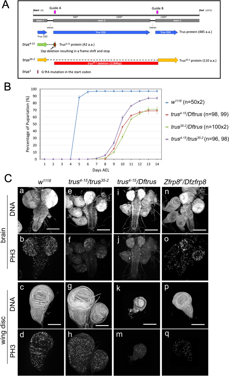
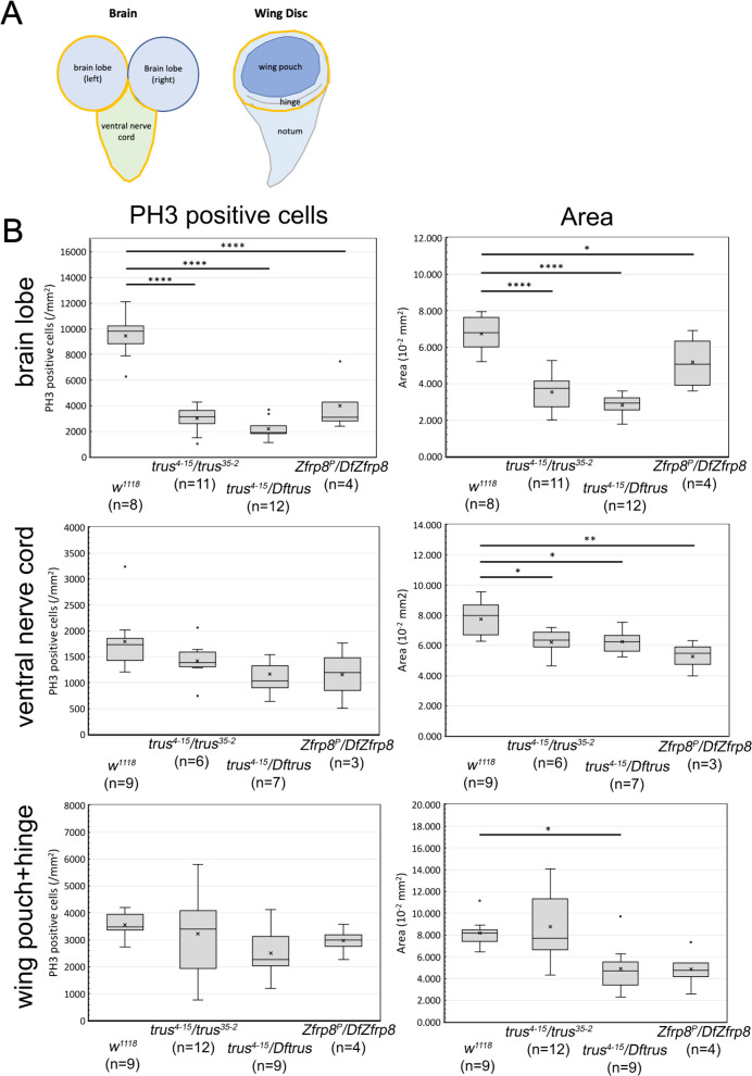
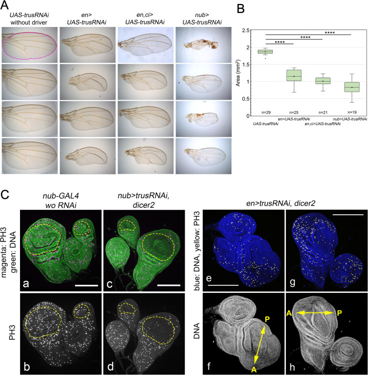
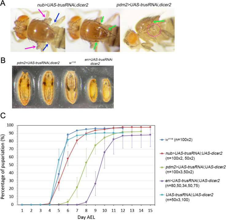
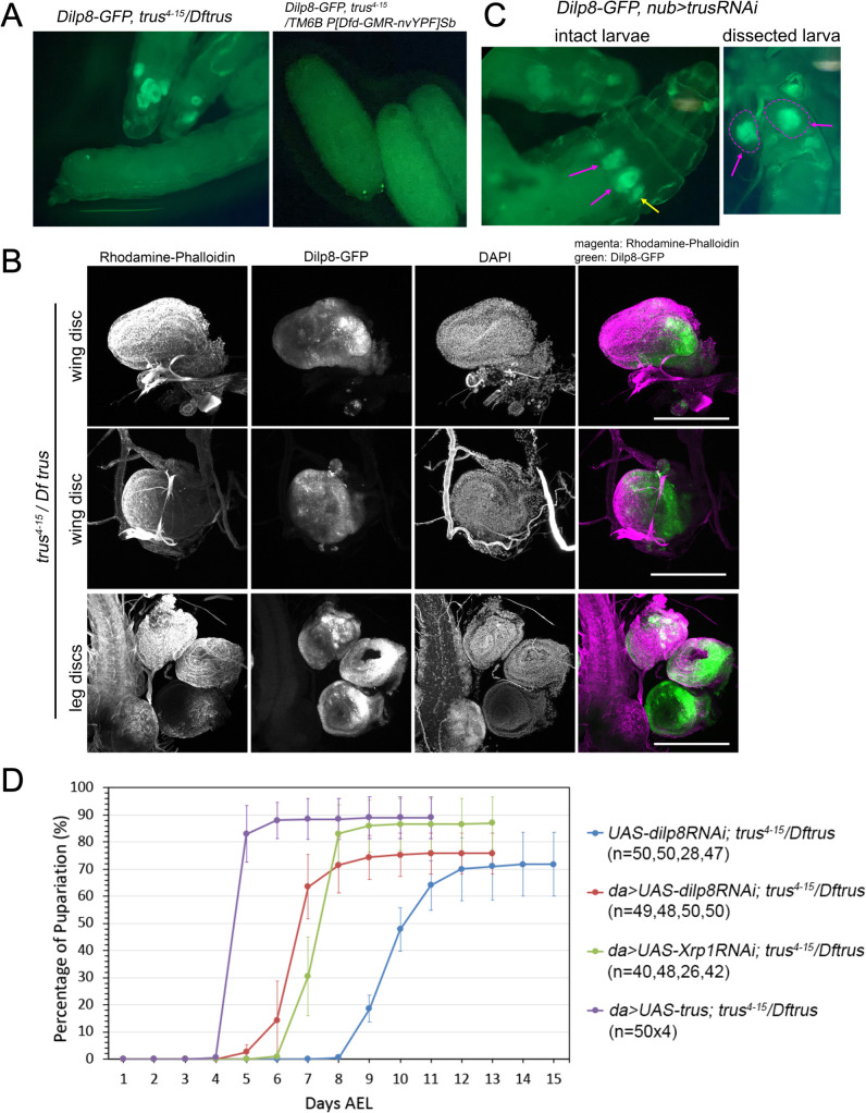
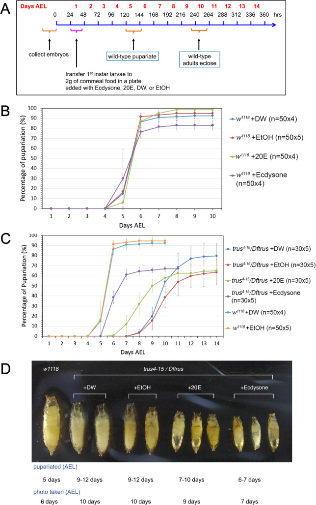
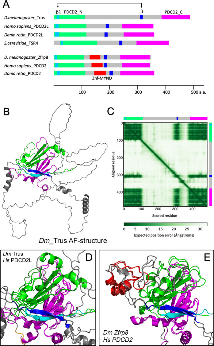
Toys are us (Trus) is the Drosophila melanogaster ortholog of mammalian Programmed Cell Death 2-Like (PDCD2L), a protein that has been implicated in ribosome biogenesis, cell cycle regulation, and oncogenesis. In this study, we examined the function of Trus during Drosophila development. CRISPR/Cas9 generated null mutations in trus lead to partial embryonic lethality, significant larval developmental delay, and complete pre-pupal lethality. In mutant larvae, we found decreased cell proliferation and growth defects in the brain and imaginal discs. Mapping relevant tissues for Trus function using trus RNAi and trus mutant rescue experiments revealed that imaginal disc defects are primarily responsible for the developmental delay, while the pre-pupal lethality is likely associated with faulty central nervous system (CNS) development. Examination of the molecular mechanism behind the developmental delay phenotype revealed that trus mutations induce the Xrp1-Dilp8 ribosomal stress-response in growth-impaired imaginal discs, and this signaling pathway attenuates production of the hormone ecdysone in the prothoracic gland. Additional Tap-tagging and mass spectrometry of components in Trus complexes isolated from Drosophila Kc cells identified Ribosomal protein subunit 2 (RpS2), which is coded by string of pearls (sop) in Drosophila, and Eukaryotic translation elongation factor 1 alpha 1 (eEF1α1) as interacting factors. We discuss the implication of these findings with respect to the similarity and differences in trus genetic null mutant phenotypes compared to the haplo-insufficiency phenotypes produced by heterozygosity for mutants in Minute genes and other genes involved in ribosome biogenesis.


Conflict of interest statement

The authors have declared that no competing interests exist.

Figures

Fig 1
Fig 1. CRISPR/Cas9 induced trus mutations cause developmental delay and defects in tissue growth and cell proliferation during the larval stage.
(A) A diagram showing trus genomic region and trus mutant alleles that are used in this study. Trus CDS is shown in blue. Two guide RNAs were designed to flank the entire exon2 and the following intron (magenta). trus4-15 has a single bp deletion at 3R:12,456,811 within target A (red). This results in a truncated 42 amino acid peptide with identity up to Arg40, two missense codons, and a premature stop (green+red+brown). Since this peptide is so small even if it is stably expressed, we consider trus4-15 to be a trus null allele. A second allele, trus35-2 is a deletion of 1184 bp between 3R:12,456,805 and 3R:12,457,988 starting 3 bp upstream of target A and ending 5 bp downstream of target B (red). This deletion might give rise to a shorter 110 amino acid peptide lacking residues between Trp38 and Phe414 if stable (orange). Since trus1 allele has a point mutation that alters the initiation codon and the mutant behaves like antimorph, it is possible that the mutation results in an aberrant translational initiation from one of the alternative initiation sites and produces a short fragment. There are two additional candidate initiation sequences which weakly align to the Kozak consensus [90,91] and are in-frame with the Trus protein sequence. Candidate 1 is from +381 bp downstream and candidate 2 is from +579 bp downstream of the original initiation start codon and could produce N-terminally truncated 238 a.a. (~27kDa) or 173 a.a. (~19kDa) Trus fragments, respectively. (B) Pupariation timing of trus mutant and w1118. Each data point on the graph indicates an average pupariation percentage of two separate plates. The vertical line on each data point indicates the standard deviation. Numbers of 1st instar larvae picked at 1 Day AEL are shown in parentheses after the genotype. Percentage of pupariation at 14 days AEL were 97%, 87%, 69%, 71% for w1118, trus4-15/trus35-2, trus4-15/Dftrus, and trus35-2/Dftrus, respectively. After pupariation, most of the trus mutant larvae did not develope further and eventually died (pre-pupal lethal) except for some trus4-15/trus35-2 that developed to pharates but did not eclose. More than 95% of the w1118 animals eclosed as adults. (C) Representative images of brains and wing discs that were dissected from third instar wandering larvae. Genotypes indicated at the top of the panels. DNA was stained with DAPI and mitotic cells are detected with anti-phospho-HistoneH3 (PH3) antibody. Maximum intensity Z-projections are shown. Scale bar: 200μm.
Fig 2
Fig 2. trus mutation caused a significant reduction of mitotic cell number and size of brain and wing disc.
(A) Diagrams showing the larval brain and wing-disc. The areas surrounded by yellow lines indicates brain lobe, ventral nerve cord, and wing pouch plus hinge areas that are quantified in B. (B) Quantification of PH3 positive cells/mm2 (left column) and area in mm2 (right column) for brain lobe, ventral nerve cord, and [wing pouch + hinge] are shown. Genotypes and number of tissues measured for each genotype are indicated in parenthesis under the graphs. The x in the boxes indicates the mean value and the line inside the box indicates the median. Box-and-whisker plots were generated using Microsoft Excel (Redmond, WA) and ANOVA analysis followed by Dunnett’s test to compare each mutant to wild-type (w1118) were performed using Prism 10 (GraphPad Software, INC., Boston MA). When the P-value in Dunnett’s test indicates that the pair variance is statistically significant, it is shown as a horizontal line across the w1118 and the mutant with * (P<=0.05), ** (P<=0.01), *** (P<=0.001), or **** (P<=0.0001) (data in S2 File). PH3 positive cells and area measurements of [wing pouch+hinge] from trus4-15/trus35-2 larvae show significant variations within the group, some wing discs are small, and others are overgrown, suggesting that the trus35-2 allele that possibly produces a 110 a.a. Trus fragment is causing hypo/hyper-morphic effects in combination with the protein null trus4-15 allele. The observed variation is not likely to be caused by experimental errors but is merely indicative of the trus4-15/trus35-2 mutant phenotype. When we removed the intrinsically variable trus4-15/trus35-2 group from ANOVA analysis of [wing pouch+hinge], the results for PH3 positive cell count and area measurement showed P < 0.05 (*) and P < 0.01 (**), respectively. Following Dunnett’s tests indicated P < 0.01 (**) for both PH3 count and area comparisons between trus4-15/Dftrus vs. w1118 (see ANOVA analysis 2 in S2 File). This indicates that wing disc growth is clearly inhibited in trus null mutant (trus4-15/Dftrus) compared to w1118. The Dunnett’s test also showed that area of [wing pouch+hinge] from Zfrp8P/DfZfrp8 larvae compared to w1118 was significantly smaller with P < 0.05 (*).
Fig 3
Fig 3. trusRNAi induced with various wing disc drivers affects wing size and morphology and decreases cellular proliferation.
(A) Representative images of wings from female adult flies that had Trus RNAi induced with wing disc drivers. Adult wings from flies carrying UAS-trusRNAi without any driver, en-GAL4 driven UAS-trusRNAi, en and ci-GAL4 driven UAS-trusRNAi, or nub-GAL4 driven UAS-trusRNAi are shown. (B) Quantification of wing area calculated in mm2. An example area is represented as magenta dashed outline in the top-left in A. ImageJ/Fiji (https://imagej.net) was used for area measurement. Box-and-whisker plots were generated using Microsoft Excel. The x in the boxes indicates the mean value and the line inside the box indicates the median. Sample numbers are indicated at the bottom of the graph (ex. n = 29 for UAS-trusRNAi). ANOVA analysis followed by Dunnett’s test to compare wing area of each RNAi samples to w1118 were performed using Prism 10 (GraphPad Software, INC.). **** indicates P value <=0.0001 (S3 File). (C) Reduction of anti-PH3 stained foci is observed in the pouch area of wing and haltere discs in nub>trusRNAi, UAS-dicer2 larvae (a-d) and the posterior half of wing discs and one half of the leg disc in en>trusRNAi, UAS-dicer2 larvae (e-h). In a and c, green: DAPI staining and magenta: anti-PH3 staining. Yellow dashed lines indicate pouch of wing and haltere discs. In e and g, blue: DAPI staining and yellow: anti-PH3 staining. Arrows in f and h indicate the anterior (A)- posterior (P) axis of wing discs. For each genotype, more than 12 wing discs were dissected and they showed the similar phenotypes. Maximum intensity Z-projections are shown. Bar: 200μm.
Fig 4
Fig 4. trusRNAi induced with wing disc drivers delay pupariation timing.
(A) trusRNAi induced with either nub-GAL4 or pdm2-GAL4 in the presence of UAS-dicer2 show a complete loss of wing blade (magenta arrows and magenta dotted-circle), morphological defects in halteres (blue arrows), and extra/disorganized bristles (green arrows). (B) (left) pdm2 > trusRNAi pharates with wing defects. (right) en>trusRNAi larvae pupariate precociously resulting in smaller pre-pupae that never become pupae. (middle) w1118 (control) pupa shown for size and structural comparison. (C) Pupariation timing of trusRNAi larvae induced with nub-GAL4 (red), pdm2-GAL4 (green), or en-GAL4 (purple). Pupariation timing of w1118 larvae (blue) and larvae carrying UAS-trusRNAi and UAS-dicer2 without driver (light blue) are shown as controls. Each data point represents an average pupariation percentage from multiple plates. The vertical line on each data point indicates the standard deviation. Numbers of 1st instar larvae picked at 1 Day AEL are shown in parentheses after the genotype.
Fig 5
Fig 5. Xrp1-Dilp8 pathway is activated leading to developmental delay in trus mutants.
(A) (left) Dilp8-GFP expression in third instar wandering trus 4-15/ Dftrus larvae. (right) Dilp8-GFP expression in 3rd instar wandering trus4-15/ TM6B P[Dfd-GMR-nvYPF]Sb or Dftrus/ TM6B P[Dfd-GMR-nvYPF]Sb larvae. Two bright GFP dots are Dfd-GMR-nvYFP signals on eyes. (B) Fixed and dissected wing (first and second rows) and leg (bottom row) discs from trus 4-15/ Dftrus third instar wandering larvae show Dilp8-GFP expression (green). Rhodamine-Phalloidin and DAPI staining reveal significant reduction in size and abnormal morphologies of the discs. 16 larvae were dissected, and representative images are shown. Maximum intensity Z-projections are shown. Scale bar: 200μm. (C) (left) Expression of Dilp8-GFP in the pouch region of the wing disc (magenta arrows) and haltere disc (yellow arrow) from nub> trusRNAi larvae. (right) A dissected nub>trus RNAi larval wing discs are marked with a magenta dashed line and show GFP fluorescence in the middle area of the wing discs (wing pouch). (D) Developmental timing curves show that da>dilp8RNAi (red) or da > Xrp1RNAi (green) significantly rescue the developmental delay of trus4-15/Dftrus larvae. da > UAS-trus rescues the developmental delay and lethality of trus4-15/Dftrus to the wild-type level and is shown as the positive control (purple). trus4-15/Dftrus larvae with the UAS-dilp8RNAi transgene without driver serve as the negative control (blue).
Fig 6
Fig 6. Ecdysone feeding to trus mutant larvae accelerates pupariation timing but causes precocious pupariation and does not rescue the pre-pupal lethality.
(A) Schedule of the ecdysone-feeding experiment. The larvae were fed mashed regular cornmeal fly food that was mixed with 20E or ecdysone dissolved in solvent (ethanol) beginning at the 1st instar stage (1 day AEL). For the controls, fly food was mixed with either water or ethanol only. (B) Pupariation timing of w1118 larvae fed cornmeal fly food mixed with either ecdysone (purple), 20-hydroxyecdysone (20E) (green), dH2O (blue), or ethanol (red). (C) Pupariation timing of trus4-15/Dftrus larvae fed cornmeal food mixed with either ecdysone (purple), 20E (green), dH2O (blue), or ethanol (red). Pupariation timing of w1118 fed cornmeal food mixed with dH2O (light blue), or ethanol (orange) are from the same data set shown in B. Each circle represents the average percentage of pupariated larvae from multiple dishes of the same genotype. Pupariated larvae were counted every 24 hours. Standard deviations are shown as vertical lines for each data point in B and C. (D) Pre-pupae that were fed cornmeal food mixed with either dH2O, ethanol, 20E, or ecdysone with the time of pupariation indicated below. w1118 pupa is shown on the left for size comparison.
Fig 7
Fig 7. Predicted 3D Structure of Trus and its paralog Zfrp8 share a core module that is conserved through evolution.
(A) Domain structure comparison of Drosophila Trus, its paralog Zfrp8, and their orthologs from different organisms. D. melanogaster Trus (Accession number: Q9VG62; Dmel\CG5333), Homo sapiens_PDCD2L (Q9BRP1), Danio retio_PDCD2L (Q5RGB3), S. cerevisiae_Tsr4 (P87156), D. melanogaster_Zfrp8 (Q9W1A3), Homo sapiens_PDCD2 (Q16342), and Danio retio_PDCD2 (Q1MTH6) are shown. Green: PDCD2_N, magenta: PDCD2_C, red: MYND-type zinc finger domain (Znf-MYND), light blue: first β-strand in PDCD2_N domain, blue: β-strand that is predicted to interact with the first β-strand, gray: unstructured loop. (B) D. melanogaster Trus protein 3D structure predicted by AlphaFold (https://alphafold.ebi.ac.uk/). Color-coding of domains are the same as shown in A. The PDCD2_N consists of four β-strands from which three β-strands form a β-sheet structure immediately followed by an α-helix (shown in green). In addition, the first β-strand in the PDCD2_N domain (residues 9-16, shown in light blue) is predicted to form hydrogen bonds with another β-strand (residues 307-317, shown in blue) in the middle of the loop region between the domain PDCD2_N and the domain PDCD2_C. (C) Predicted Aligned Error (PAE) of Drosophila Trus 3-D structure calculated by the AlphaFold. Color-coded bars representing the Trus protein domains shown in B are placed on upper and right sides of the panel. The PAE indicates high confidence in the relative position of scored residues 1-109 (PDCD2_N; shown in light blue and green) when aligned with residues 382-485 (PDCD2_C; shown in magenta), supporting the packing between these regions that form a structural module despite the large unstructured loops (gray) that separate the PDCD2_N and PDCD2_C domains. The PAE of the blue β−strands (residues 307-317) against both the N-terminal domain (PDCD2_N, residues 1-109) and the C-terminal domain (PDCD2_C, residues 382-485) shows high confidence, indicating packing of a core module that includes the PDCD2_N (light blue and green, residues 1-109), the PDCD2_C (magenta, residue 382-485), and a β-strand in-between (blue, residues 307-317). (D) Structural alignment of Dm Trus and Hs PDCD2L shows evolutional conservation of the core module in the PDCD2L protein family. (E) Structural alignment of Dm Zfrp9 and Hs PDCD2 shows evolutional conservation of the core module in the PDCD2 protein family. 3-D structural alignments were performed with PyMOL (Schrödinger LLC., NY).
Fig 8
Fig 8. trus expresses in mitotic tissues.
In situ hybridization with anti-sense RNA that hybridizes with trus mRNA reveals high level expression of trus in larval lymph gland, ovary, wing disc, gut, and brain lobe. Low expression is detected in ring and salivary glands.
Fig 9
Fig 9. Trus localizes to the cytoplasm in cultured cells and in vivo.
(A) Drosophila Trus localizes to the cytoplasm in S2 cells and shuttles between the nucleus and the cytoplasm in a CRM1/XPO1-dependent manner. Leptomycin B (LMB) is an inhibitor of CRM1/XPO1. Without LMB treatment (top row), EGFP-Trus expressed in S2 cells localizes primarily to the cytoplasm, and after treatment of the cells with LMB (middle and bottom rows), EGFP-Trus accumulates in the nucleus (LMB 15min, LMB 115min). Maximum intensity Z-projections are shown. Bar: 20μm. (B) da-GAL4 induced UAS-EGFP-trus expression in larval tissues. da-GAL4 expression is ubiquitous in most of the tissues. EGFP-Trus primarily localizes in the cytoplasm of cells (b, f, and j). Representative images of a wing disc (a-d), brain (e-h), and prothoracic grand (i-k) are shown. (b-d) and (f-h) are magnified images of the area marked with a yellow square in a and e, respectively. Each image is a single Z slice from multiple Z series confocal scanning data. Scale bar: 200μm.
Fig 10
Fig 10. Rescue of lethality and developmental delay of the trus mutant can be achieved by Trus expression induced with specific drivers.
(A) Developmental timing of trus mutants (trus4-15/Dftrus) with trus expression driven by various drivers display varying degrees of rescue. Compared to the w1118 control (orange), da-Gal4 (dark blue) rescues the best, with en-Gal4 and ci-Gal4 (aqua), en-Gal4 (green), and ci-Gal4 (purple) showing progressively lower degrees of rescue. ci-Gal4 without UAS-trus (red) serves as the negative control. (B) Adult and pupal phenotypes of rescued lines. ci > UAS-trus rescued trus4-15/Dftrus pre-pupal lethality with significant defects in wings, halteres, legs, and bristles (upper panels). Many flies eclose only half-way from the pupal case and die (lower left). trus4-15/Dftrus mutants without the UAS-trus transgene arrest and die during the pre-pupal stage (lower right). (C) (a-f, c’, and f’) Ovariole phenotypes of rescued lines confirms that da > UAS-trus rescue of trus4-15/Dftrus mutant females. They are fertile and show no defects in oogenesis. The yellow star in d indicates a mature egg produced. (g-m, i’, and m’) en,ci > UAS-trus in combination with trus4-15/Dftrus mutants, while being able to rescue mutant lethality, give rise to females that are sterile. Their ovarioles produce no mature eggs because egg chambers degrade at mid-oogenesis. (green) Rhodamine-Phalloidin, (blue) DAPI. For each genotype, 10 ovaries from 10 individual female were dissected and all showed the similar phenotypes. Maximum intensity Z-projections are shown. Scale bar: 200μm.

Update of

Similar articles

References

    1. Parker MD, Karbstein K. Quality control ensures fidelity in ribosome assembly and cellular health. J Cell Biol. 2023;222(4):e202209115. doi: 10.1083/jcb.202209115 - DOI - PMC - PubMed
    1. Ni C, Buszczak M. Ribosome biogenesis and function in development and disease. Development. 2023;150(5):dev201187. doi: 10.1242/dev.201187 - DOI - PMC - PubMed
    1. Bridges C, Morgan T. The third-chromosome group of mutant characters of Drosophila melanogaster. Carnegie Institute of Washington. 1923.
    1. Kongsuwan K, Yu Q, Vincent A, Frisardi MC, Rosbash M, Lengyel JA, et al. A Drosophila Minute gene encodes a ribosomal protein. Nature. 1985;317(6037):555–8. doi: 10.1038/317555a0 - DOI - PubMed
    1. Andersson S, Saebøe-Larssen S, Lambertsson A, Merriam J, Jacobs-Lorena M. A Drosophila third chromosome Minute locus encodes a ribosomal protein. Genetics. 1994;137(2):513–20. doi: 10.1093/genetics/137.2.513 - DOI - PMC - PubMed

MeSH terms

LinkOut - more resources