. 2025 Jun 27;21(6):e1011750.
doi: 10.1371/journal.pgen.1011750. eCollection 2025 Jun.

Functional analysis of CYP4B1 enzymes from apes and humans uncovers evolutionary hot spots for adaptations of the catalytical function

Affiliations

Functional analysis of CYP4B1 enzymes from apes and humans uncovers evolutionary hot spots for adaptations of the catalytical function

Saskia Hüsken et al. PLoS Genet. .

Abstract

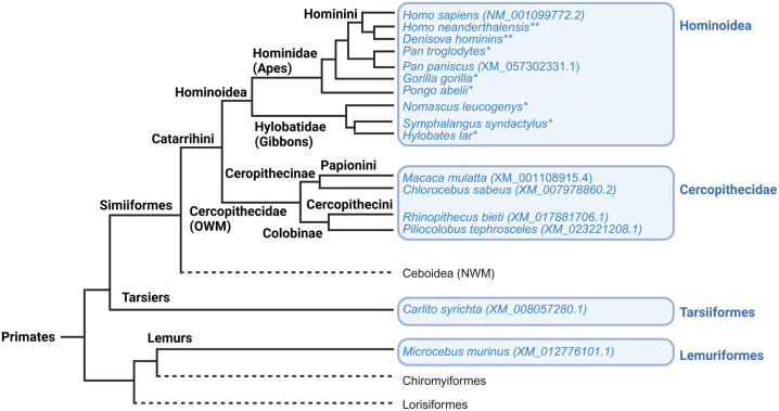
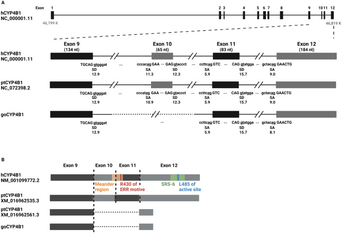
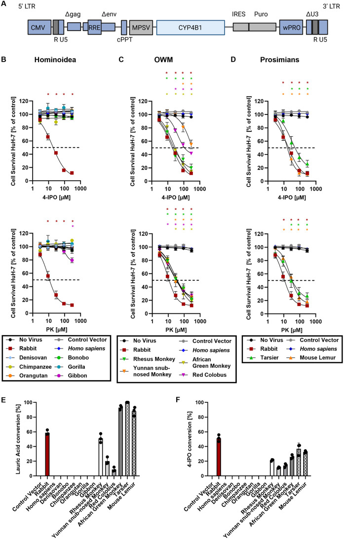
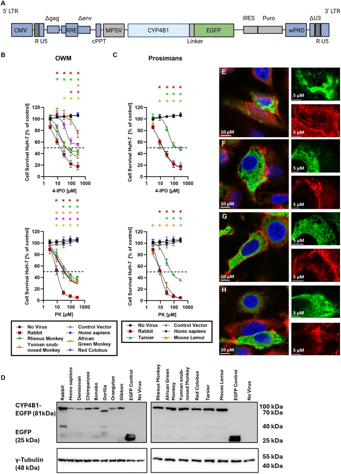
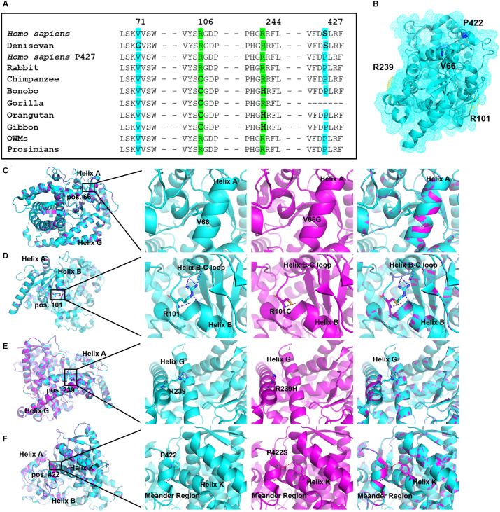
A hallmark of the highly conserved CYP4B1 enzyme in mammals is the capability to bioactivate both xenobiotic and endobiotic substrates. However, due to a single amino acid change (p.P427S) within the evolutionary conserved meander region no catalytic activity of the native human CYP4B1 has been identified so far. To identify at which point in human evolution the loss of CYP4B1 activity had occurred, we evaluated the activities of CYP4B1 orthologs from 14 primate genera against 4-ipomeanol and perilla ketone in human liver cells. The activity of recombinant CYP4B1 proteins isolated from E. coli was also tested against 4-ipomeanol and lauric acid. Surprisingly, CYP4B1 already became catalytically inactive at the split between apes and monkeys; all tested CYP4B1 orthologs from monkeys were able to bioactivate both protoxins and to hydroxylate lauric acid. Amino acid analysis of the CYP4B1 orthologs revealed four additional evolutionary changes, each affecting the function of ape and human enzymes: p.V71G specific for Denisovans, p.R106C, p.R244H, and an exon deletion found only in the gorilla CYP4B1. Systematic functional analyses proved the negative impact of the genetic changes on CYP4B1 activity and showed that reversion of the mutations restored enzyme activity. The occurrence of five independent inactivating genetic changes in the same gene of closely related species is a clear indication of the importance of inactivating CYP4B1 in apes and humans. Elucidating the evolutionary trigger(s) for CYP4B1 inactivation in our ancestors will ultimately improve our understanding of primate evolution.


Conflict of interest statement

The authors have declared that no competing interests exist.

Figures

Fig 1
Fig 1. Schematic primate phylogeny based on whole-genome sequences of primates (compiled from [–39]).
CYP4B1 sequences from species marked in blue were used in this study for CYP4B1 characterization. Sequences were either downloaded from NCBI, provided by the MPI, Leipzig (**), or curated by our team (*) after sequencing of RNA extracted from cryopreserved lung tissues.
Fig 2
Fig 2. Sequence analysis of gorilla CYP4B1.
(A) The endogenous Homo sapiens CYP4B1 (hCYP4B1) located on human chromosome 1 (NC_000001.11 NCBI ID) encodes 12 exons. (B) gDNA sequence analysis of three gorilla lung tissues samples revealed a gap spanning part of intron 9, exon 10, and intron 10 when compared to hCYP4B1 and chimpanzee CYP4B1 (ptCYP4B1, NC_072398.2 NCBI-ID). The strengths/scores of the splice acceptor (SA) and splice donor (SD) sites were calculated using MAXENT or HBS splice site algorithms. At the mRNA level, gorilla CYP4B1 (goCYP4B1) is lacking not only exon 10, but also exon 11 as shown by mRNA sequencing of lung tissues. Functional domains highlighted as follows: substrate recognition sites (green), heme covalent binding site (yellow), the ERR triad (red), active site (blue), and meander region (orange).
Fig 3
Fig 3. Functional analysis of primate CYP4B1 orthologs.
(A) The cDNAs of CYP4B1 enzymes from different primate clades, including prosimians, Old World monkeys (OWMs), gibbon, great apes, and humans were cloned into a lentiviral expressing vector upstream of an IRES-Puro site. CMV: CMV promoter; LTR: long terminal repeat; RRE: Rev responsive element, cPPT: central polypurine binding tract; MPSV: U3 promoter of the myeloproliferative sarcoma virus; IRES: internal ribosomal entry site; Puro: cDNA for puromycin resistance; WPRO: woodchuck hepatitis virus post-transcriptional regulatory element optimized. (B-D) The activities of CYP4B1 orthologs in HuH-7 liver cells were evaluated in MTS assays following a 24 hrs exposure to either 4-ipomeanol (4-IPO, upper graphs) or Perilla ketone (PK, lower graphs). Experiments were performed with a minimum of three individual replicates each. The graphs are divided based on primate lineages: gibbon and great apes (B), Old World monkeys (C), and prosimians (D). (E-F) Specific activities for hydroxylation of LA and metabolization 4-IPO by recombinant CYP4B1 orthologs isolated from E. coli. For each data set at least three individual replicates were measured and are shown as mean ± SEM. For statistical analysis, a multiple comparison one-way ANOVA with subsequent Dunnett’s-Post-hoc test was used to determine significant differences between measuring points compared to untreated controls: p-values < 0.05 were defined as significant and were marked with an asterix (*). The underlying data for the graphs in this figure can be found in S1 Data. For calculation of the half-maximal effective concentration that reduces the surviving cell number by 50% (GI50 values), a non-linear fit model was applied to each data set (S3 Data).
Fig 4
Fig 4. Activity, expression and localization of CYP4B1-EGFP fusion proteins in HuH-7 cells.
(A) Design of the lentiviral vector used for overexpression of the CYP4B1-EGFP fusion proteins. (B-C) Enzymatic activities and of the fusion proteins of Old World monkeys (OWMs) and prosimians against 4-IPO and PK in HuH-7 cells. GI50 values calculated based on the non-linear cur fit model can be found in S3 Data. (D) Expression levels of CYP4B1-EGFP fusion proteins in polyclonal HuH-7 cell cultures were estimated by western blot. For detection, an anti-EGFP antibody was used; γ-Tubulin was used as a loading control. (E-H) Subcellular localization of the fusion proteins of OWMs and prosimians. Shown are representative images of cells overexpressing either rabbit (E), mouse lemur (F), bonobo (G), or wild type Homo sapiens (H) CYP4B1. The cells were co-stained for nuclei (HOECHST) and F-actin (Phalloidin-TRITC). 10 µM or 5 µM scale bars are implemented on overview images or detailed images showing only one fluorescent channel (EGFP or phalloidin), respectively. For each data set at least three individual replicates were measured and are shown as mean ± SEM. For statistical analysis, a multiple comparison one-way ANOVA with subsequent Dunnett’s-Post-hoc test was used to determine significant differences between measuring points compared to untreated controls: p-values < 0.05 were defined as significant and were marked with an asterix (*). The underlying data for the graphs in this figure can be found in S1 Data.
Fig 5
Fig 5. In silico modeling of potential key amino acid positions for the functional activity of mammalian CYP4B1.
(A) Multiplexed comparison of CYP4B1 amino acid sequences to identify hot spots with potential impact on catalytic function. Human-based amino acid exchanges are colored in blue, while substitutions in apes are colored in green. (B) The location of all corresponding residues is depicted within the rabbit CYP4B1 model, PDB: 5T6Q (rabbit CYP4B1 complexed with octane). Within the rabbit CYP4B1, the amino acid sequence positions are shifted by -5 when compared to primate CYP4B1 sequences. Residues are colored in blue (p.V66 and p.P422) or green (p.R101 and p.R239). (C-F) Side directed mutagenesis of all four key amino acid positions (p.V66, p.R101, p.R239, and p.P422) was performed based on the rabbit CYP4B1 protein structure. Overall structure is colored in cyan (WT sequence) or magenta (mutated enzyme), amino acid residues are colored by atoms, and polar interactions are depicted as yellow dots. From left to right: location within the whole enzyme, defined WT amino acid sequence of rabbit CYP4B1, defined mutated amino acid, superimposition of WT and mutated enzyme.
Fig 6
Fig 6. Functional consequences of the four critical amino acid substitutions in Homo sapiens and rabbit CYP4B1.
All experiments were performed with a minimum of three individual replicates each in the human liver carcinoma cell line HuH-7. (A) Enzyme activity of the Homo sapiens CYP4B1 wild type protein and p.S427P variant and of the Denisovan wild type (S427, G71) and p.S427P variant against increasing concentrations of 4-IPO or PK. (B) Enzyme activity of the rabbit CYP4B1 wild type protein and the single or double mutated variants p.P422S and p.V66G. (C) Functional effects of the amino acid substitutions p.R106C and p.R244H introduced in the Homo sapiens CYP4B1 P427 enzyme. (D). Functional implications of the amino acid substitutions at the corresponding positions in the rabbit CYP4B1 wild type and p.P422S enzyme. (A-D) For each data set at least three individual replicates were measured and are shown as mean ± SEM. For statistical analysis, a multiple comparison one-way ANOVA with subsequent Dunnett’s-Post-hoc test was used to determine significant differences between measuring points compared to untreated controls: p-values < 0.05 were defined as significant and were marked with an asterix (*). The GI50 values for the respective Homo sapiens and rabbit enzymes were calculated based on a non-linear curve fit model and can be found in S3 Data. The underlying data for the graphs in this figure can be found in S1 Data. (E). Effect of the amino acid exchanges p.P427S, p.V71G, p.R106C and p.R244H on the expression of Homo sapiens and rabbit (wild type and p.P422S variant) CYP4B1 in HuH-7. Human CYP4B1 enzymes were detected with an anti-human CYP4B1 polyclonal antibody. Rabbit CYP4B1 enzymes were detected with an anti-rabbit CYP4B1 polyclonal antibody. γ-tubulin served as a loading control.
Fig 7
Fig 7. Restoring the activity of gibbon and two great ape CYP4B1 orthologs by p.C106R and p.H244R substitution.
The enzymatic activity of hominoid CYP4B1 proteins was evaluated in HuH-7 cells after a 24h exposure to either 4-IPO or PK. (A) Enzymatic activity of bonobo CYP4B1 variants. (B) Catalytic activity of orangutan CYP4B1 variants. (C) Enzyme activity of gibbon CYP4B1 variants. For each data set at least three individual replicates were measured and are shown as mean ± SEM. For statistical analysis, a multiple comparison one-way ANOVA with subsequent Dunnett’s-Post-hoc test was used to determine significant differences between measuring points compared to untreated controls: p-values < 0.05 were defined as significant and were marked with an asterix (*). GI50 values calculated based on a non-linear curve fit model (S3 Data). The underlying data for the graphs in this figure can be found in S1 Data. (D). Effect of the amino acid exchanges p.C106R and p.H244R on expression levels of gibbon, orangutan, and bonobo CYP4B1 proteins in HuH-7 cells. The hominoid proteins were detected with an anti-human CYP4B1 antibody. γ-tubulin served as a loading control.
Fig 8
Fig 8. Synthesis of perilla ketone and 4-ipomeanol.
a) HNMeOMe, TBTU, HOBt, NEt3, DCM, RT; b) Mg, 1-bromo-3-methylbutane, THF, 0 °C to RT; c) Mg, O-TBDMS-4-bromo-2-butanol, THF, 0 °C to RT; d) TBAF, THF, RT.

Similar articles

References

    1. Nelson DR. The cytochrome p450 homepage. Hum Genomics. 2009;4(1):59–65. - PMC - PubMed
    1. Nelson DR. Cytochrome P450 diversity in the tree of life. Biochim Biophys Acta Proteins Proteom. 2018;1866(1):141–54. doi: 10.1016/j.bbapap.2017.05.003 - DOI - PMC - PubMed
    1. Dasgupta A. Chapter 4 - Genetic Factors Associated With Opioid Therapy and Opioid Addiction. In: Dasgupta A, editor. Fighting the Opioid Epidemic. Elsevier. 2020. p. 61–88.
    1. Machalz D, Pach S, Bermudez M, Bureik M, Wolber G. Structural insights into understudied human cytochrome P450 enzymes. Drug Discov Today. 2021;26(10):2456–64. doi: 10.1016/j.drudis.2021.06.006 - DOI - PubMed
    1. Gotoh O. Evolution of cytochrome p450 genes from the viewpoint of genome informatics. Biol Pharm Bull. 2012;35(6):812–7. doi: 10.1248/bpb.35.812 - DOI - PubMed