. 2025 Jun 27;21(6):e1013278.
doi: 10.1371/journal.ppat.1013278. eCollection 2025 Jun.

Viral hijacking of host DDX60 promotes Crimean-Congo haemorrhagic fever virus replication via G-quadruplex unwinding

Affiliations

Viral hijacking of host DDX60 promotes Crimean-Congo haemorrhagic fever virus replication via G-quadruplex unwinding

Yutong Sui et al. PLoS Pathog. .

Abstract

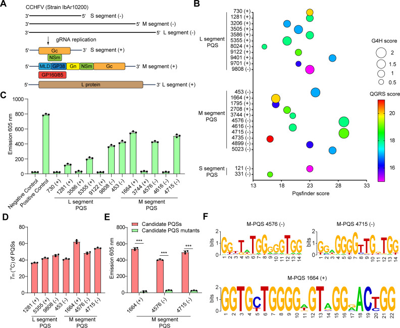
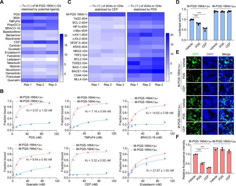
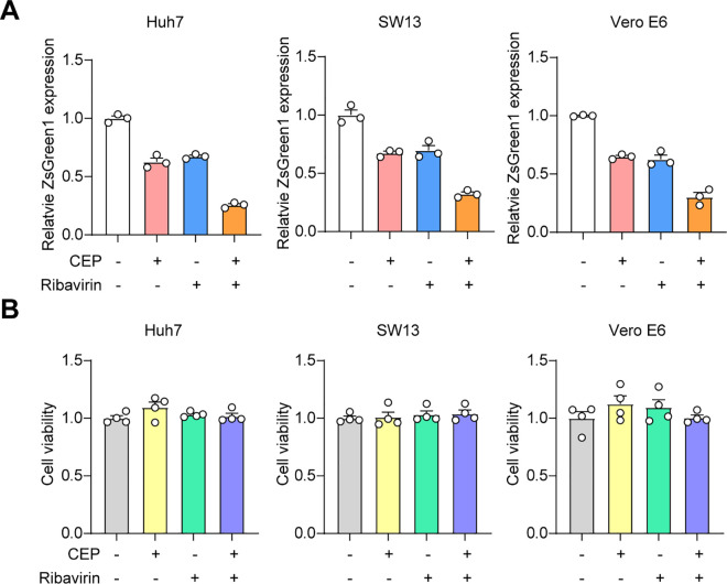
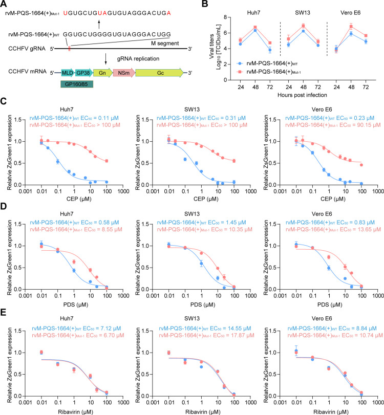
Crimean-Congo haemorrhagic fever virus (CCHFV) is the most prevalent tick-borne zoonotic bunyavirus, causing severe hemorrhagic fever and fatality in humans. Currently, the absence of approved vaccines or therapeutics for CCHFV infection necessitates the development of innovative therapeutic strategies. Here, we identify a guanine (G)-rich sequence located within the mRNA of the glycoprotein precursor in the medium (M) segment of the CCHFV genome, designated as M-PQS-1664(+). M-PQS-1664(+) can form stable G-quadruplex (G4) structure and functions as a negative regulatory element for viral replication. Host DDX60 is up-regulated in response to CCHFV infection, thereby it is hijacked to unwind M-PQS-1664(+) G4 for facilitating viral replication. The FDA-approved drug Cepharanthine (CEP), which competes with DDX60 to specifically stabilize M-PQS-1664(+) G4 without a global induction of host cellular G4s formation, exhibits remarkable antiviral activity in vitro and in vivo. More importantly, CEP possesses antiviral activity (50% inhibitory concentration ~ 0.2 μM) that having ~ 88 × the potency of ribavirin. Our findings underscore the CCHFV G4s as a promising target for drug development and highlight the significant potential of CEP in combating CCHFV.


Conflict of interest statement

The authors have declared that no competing interests exist.

Figures

Fig 1
Fig 1. Detection and annotation of G-quadruplexes (G4s) in CCHFV genome.
(A) The tri-segmented viral genome (L, M and S segments) of Crimean-Congo haemorrhagic fever virus (CCHFV) genome. L, M and S segments, large, medium and small segments. (B) CCHFV G4-forming sequences (PQSs) in the CCHFV three genomic segments. Prediction results are shown with multi-parameter analysis of Pqsfinder, G4H and QGRS scores. The (-) symbol represents CCHFV genomic RNA, while the (+) symbol denotes CCHFV mRNA. The number indicates the position of PQS within the genomic RNA of CCHFV. (C) NMM fluorescence turn-on assays for CCHFV PQS candidates. (D) Tm of CCHFV PQS candidates. (E) NMM fluorescence turn-on assays for CCHFV PQSs and their mutants. (F) The analysis of conservation of the CCHFV PQS candidates across strains. The mean of triplicate wells is represented by each point, with error bars indicating the standard error of mean (SEM). The graphs presented here are representative of three independent experiments. ***P < 0.001 by Student’s t test.
Fig 2
Fig 2. G4-specific ligands inhibits report genes expression by stabilizing CCHFV G4.
The heat map of △Tm (°C) of M-PQS-1664(+) G4 stabilized by potential ligands. (B) Binding curves of compounds and M-PQS-1664(+) G4 determined by MicroScale Thermophoresis (MST) assays, and dissociation constants (Kd) obtained from curve fitting (1:1) in the presence of G4. Error bars represent the SEM calculated from three replicates. (C) The heat map of △Tm (°C) of M-PQS-1664(+) G4, cellular dG4s or rG4s stabilized by CEP and PDS. (D) The luciferase activity in HEK293T cells transfected with luciferase vectors harboring M-PQS-1664(+)WT or M-PQS-1664(+)Mut was detected after 500 nM CEP or 500 nM PDS treatment for 24 hours by performing luciferase reporter assays. Error bars represent the SEM calculated from four replicates. (E) HEK293T cells transfected with pLV-EGFP-N vectors harboring M-PQS-1664(+)WT or M-PQS-1664(+)Mut were treated with 500 nM PDS or 500 nM CEP for 48 hours. Representative confocal images were demonstrated. Scale bars: 10 μm. (F) The relative fluorescent value of EGFP in transfected and treated HEK293T cells was measured. The mean of triplicate wells is represented by each point, with error bars indicating the SEM. The graphs presented here are representative of three independent experiments. ***P < 0.001 by Student’s t test.
Fig 3
Fig 3. DDX60 promotes CCHFV infection by unfolding G4.
Mass spectrometry representation for RNA pull-down from Huh7 cell lysates using M-PQS-1664(+) G4 and its mutated counterparts. A list of enriched proteins is mentioned showing interaction (Figure was modified from https://openclipart.org/.). (B) RNA pull-down of M-PQS-1664(+) G4, and their mutated counterparts followed by western blotting assays to detect DDX60 protein. (C) Electrophoretic mobility shift assay (EMSA) results show the binding of DDX60 to M-PQS-1664(+) G4 and Telomere G4 (TelG4). (D) DDX60 can unfold M-PQS-1664(+) G4 in concentration-dependent manners in the presence of 1 mM ATP. (E) Binding curves of DDX60 and M-PQS-1664(+) G4 determined by MST assays, and dissociation constants (Kd) obtained from curve fitting (1:1) in the presence of G4. Error bars represent the SEM calculated from three replicates. (F) The activity luciferase in DDX60-OE HEK293T cells transfected with luciferase vectors harboring M-PQS-1664(+) G4WT or M-PQS-1664(+) G4Mut was detected by performing luciferase reporter assays. DDX60 overexpression, DDX60-OE. Error bars represent the SEM calculated from four replicates. (G) 500 nM CEP and 500 nM PDS treatment for 24 hours inhibit DDX60 mediated increase of luciferase activity. Error bars represent the SEM calculated from four replicates. (H and I) The protein levels of DDX60 in DDX60 WT (Ctrl) and DDX60 knockout (DDX60-KO) Huh7 and HepG2 cells as well as DDX60-KO Huh7 and HepG2 cells transiently transfecting DDX60. The cells were infected without or with CCHFV/ZsG at MOI 0.1., subsequently the protein levels of DDX60 (H) were detected at 72 hours post infection by western blotting assays. The ZsG fluorescence (green) was determined at 72 hours post infection (I). DDX60 knockout, DDX60-KO. (J) 500 nM PDS or 500 nM CEP treatment inhibit the interaction between DDX60 and the CCHFV RNA containing M-PQS-1664(+) in the recombinant CCHFV-infected Huh7 cells. The cells were infected with CCHFV/ZsG at MOI 0.1., and the enrichment of CCHFV RNA containing M-PQS-1664(+) by anti-DDX60 was determined at 48 hours post infection by performing RNA immunoprecipitation (RIP) assays. The mean of triplicate wells is represented by each point, with error bars indicating the SEM. The graphs presented here are representative of three independent experiments. **P < 0.01, ***P < 0.001 by Student’s t test.
Fig 4
Fig 4. Anti-CCHFV activity of candidate drugs in cell-based assays.
(A-C) Dose-response curves were generated in Huh7, SW13, or Vero E6 cells following treatment with varying concentrations of CEP (A), PDS (B) or Ribavirin (C) prior to infection with CCHFV/ZsG at MOI 0.1. The reduction in ZsG fluorescence (green) was determined at 72 hours post infection. Cell viability was determined using the same concentrations of compounds. The mean of triplicate wells is represented by each point, with error bars indicating the SEM. The graphs presented here are representative of three independent experiments. EC50, half-maximal effective doses. CC50, half-cytotoxic concentration. SI, selectivity index.
Fig 5
Fig 5. CEP combined with ribavirin inhibits CCHFV infection.
(A) The Huh7, SW13, and Vero E6 cells were treated with the indicated compounds either individually or in combination at the following concentrations: CEP (400 nM) and ribavirin (10 μM). Subsequently, the cells were infected with CCHFV/ZsG at MOI 0.1. The reduction in ZsG fluorescence (green) was determined at 72 hours post infection. Each data point represents the mean value obtained from triplicate wells, with error bars indicating standard deviation. (B) Cell viability was determined concurrently using the indicated compounds in Huh7, SW13, and Vero E6 cells. Each data point represents the mean value obtained from quadruplicate wells, with error bars indicating SEM.
Fig 6
Fig 6. CEP and PDS inhibit CCHFV infection by targeting G4.
Schematic representation of G4-disruptive mutations in the CCHFV genome. (B) Viral growth curves in Huh7, SW13, and Vero E6 cells infected with either rvM-PQS-1664(+)WT or rvM-PQS-1664(+)Mut-1 at MOI 0.1. Viral titers were determined at 24, 48, and 72 hours post infection. rvM-PQS-1664(+)WT, recombinant virus with M-PQS-1664(+)WT. rvM-PQS-1664(+)Mut-1, recombinant virus with M-PQS-1664(+)Mut-1. (C-E) Dose-response curves were generated in Huh7, SW13, or Vero E6 cells following treatment with varying concentrations of CEP (C), PDS (D) or Ribavirin (E) prior to infection with rvM-PQS-1664(+)WT or rvM-PQS-1664(+)Mut-1 at MOI 0.1. The reduction in ZsG fluorescence (green) was determined at 72 hours post infection. The mean of triplicate wells is represented by each point, with error bars indicating the SEM. The graphs presented here are representative of three independent experiments.
Fig 7
Fig 7. CEP protects mice from CCHFV pathogenesis by targeting G4.
(A and B) Protective effects of CEP (25 mg/kg) on fatality caused by rvM-PQS-1664(+)WT or rvM-PQS-1664(+)Mut-1 infection. Body weight (A) and survival (B) of the mice (n = 7) were monitored daily. (C and D) Protective effects of CEP (25 mg/kg) on rvM-PQS-1664(+)WT or rvM-PQS-1664(+)Mut-1 caused pathogenesis. Viral cloads were measured using qRT-PCR and expressed as the number of viral RNA copies per microgram of tissue or per mL of serum (C). Hematoxylin and eosin (H&E) staining was conducted to demonstrate the pathological alterations. (D). The extensive necrosis (black asterisks) and necrotic cellular debris (black arrows) in liver were indicated. Scale bars: 100 μm. n.s., no significance. *P < 0.05, **P < 0.01 by Student’s t test.

Similar articles

References

    1. Hawman DW, Feldmann H. Crimean-Congo haemorrhagic fever virus. Nat Rev Microbiol. 2023;21(7):463–77. doi: 10.1038/s41579-023-00871-9 - DOI - PMC - PubMed
    1. Messina JP, Pigott DM, Golding N, Duda KA, Brownstein JS. The global distribution of Crimean-Congo hemorrhagic fever. Trans R Soc Trop Med Hyg. 2015;109:503–13. - PMC - PubMed
    1. Al-Abri SS, Abaidani IA, Fazlalipour M, Mostafavi E, Leblebicioglu H, Pshenichnaya N, et al. Current status of Crimean-Congo haemorrhagic fever in the World Health Organization Eastern Mediterranean Region: issues, challenges, and future directions. Int J Infect Dis. 2017;58:82–9. doi: 10.1016/j.ijid.2017.02.018 - DOI - PMC - PubMed
    1. Frank MG, Weaver G, Raabe V; State of the Clinical Science Working Group of the National Emerging Pathogens T, Education Center’s Special Pathogens Research N. Crimean-Congo Hemorrhagic Fever Virus for Clinicians-Epidemiology, Clinical Manifestations, and Prevention. Emerg Infect Dis. 2024;30:854–63. - PMC - PubMed
    1. Dokuzoguz B, Celikbas AK, Gök ŞE, Baykam N, Eroglu MN, Ergönül Ö. Severity scoring index for Crimean-Congo hemorrhagic fever and the impact of ribavirin and corticosteroids on fatality. Clin Infect Dis. 2013;57(9):1270–4. doi: 10.1093/cid/cit527 - DOI - PubMed

MeSH terms

LinkOut - more resources