. 2025 Jun 27;11(26):eadt6195.
doi: 10.1126/sciadv.adt6195. Epub 2025 Jun 27.

Hepatic NMNAT1 is required to defend against alcohol-associated fatty liver disease

Affiliations

Hepatic NMNAT1 is required to defend against alcohol-associated fatty liver disease

Qinchao Ding et al. Sci Adv. .

Abstract

Nicotinamide mononucleotide adenylyltransferase 1 (NMNAT1), a nicotinamide adenine dinucleotide (NAD+) synthetase in Preiss-Handler and salvage pathways, governs nuclear NAD+ homeostasis. This study investigated the role of NMNAT1 in alcohol-associated liver disease (ALD). Decreased NMNAT1 expression and activity were observed in the liver of patients with alcohol-associated hepatitis and either liver or primary hepatocytes from ALD mice. F-box and WD repeat domain containing 7 (FBXW7)-regulated interferon regulatory factor 1 (IRF1) ubiquitination degradation contributed to the alcohol-inhibited NMNAT1 transcriptional level. Hepatic NMNAT1 knockout aggravated alcohol-induced hepatic NAD+ decline and further hepatic steatosis and liver injury. Metabolomics and transcriptomics interaction revealed that the cysteine sulfinic acid decarboxylase (CSAD)-regulated taurine pathway was involved in NMNAT1-disrupted hepatic lipid metabolism in ALD. Hepatic CSAD overexpression or taurine supply attenuated hepatic NMNAT1 knockout-aggravated ALD. Hepatic NMNAT1 loss inhibited NMN-protected ALD. Replenishing hepatic NMNAT1 reversed liver lipid accumulation in ALD mice. These findings identified NMNAT1 as a promising therapeutic target for ALD.


Figures

Fig. 1.
Fig. 1.. Hepatic NMNAT1 is suppressed in both humans and mice with ALD.
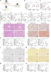
(A) Schematic diagram of the NAD+ synthesis pathway. (B to H) Representative immunohistochemical images of hepatic NMANT1 (B), NMNAT1 protein expression (C), Nmnat1 mRNA expression (D), nuclear NAD+ content (E), nuclear NADH content (F), nuclear NAD+/NADH ratio (G), and NMNAT1 activity (H) were obtained in the Lieber-DeCarli diet plus single binge model (n = 4 to 8). (I to K) Liver NMNAT1 protein expression (I), mRNA expression (J), and nuclear NAD+ content (K) were detected at different stages of Lieber-DeCarli alcohol feeding (n = 4 to 8). (L) Correlation analysis of Nmnat1 mRNA expression with plasma ALT and AST and liver TG (n = 32). (M) NMNAT1 protein expression in patients with AH (n = 5) and healthy individuals (n = 5). (N to S) Primary hepatocyte nuclei were isolated from the livers of Lieber-DeCarli diet–fed PF and AF mice, and NMNAT1 protein expression (N) and mRNA expression (O), as well as NAD+ (P), NADH (Q), NAD+/NADH ratio (R), and NMNAT1 activity (S), were detected (n = 3). The protein band intensity was quantified by ImageJ. Data are presented as the means ± SD. *P < 0.05 represents statistical difference.
Fig. 2.
Fig. 2.. Alcohol-stimulated IRF1 ubiquitination degradation contributes to NMNAT1 reduction.
(A) Venn plot of nuclear transcription factor prediction for NMNAT1. (B) Relative expression of Irf1, Rarg, and Mitf in the AF mouse liver (n = 8). (C) IRF1, RARG, and MITF protein expressions in the AF mouse liver (n = 4). (D) IRF1 protein expression in patients with AH (n = 5) and healthy individuals (n = 4). (E) Primary hepatocyte nuclei were isolated from the livers of Lieber-DeCarli diet–fed PF and AF mice. IRF1 protein expression was detected (n = 4). (F and G) AML-12 cells were treated with/without IFN-γ (1 ng/ml) for 6 hours, and IRF1 recruitment was determined by ChIP assay. DNA was extracted from the immunoprecipitates, and a fragment of the ribosomal DNA (rDNA) promoter sequence was amplified by real-time qPCR. PCR products were analyzed by 2% agarose gel electrophoresis (F). The rDNA levels were quantified by real-time qPCR [(G); n = 3]. IgG, immunoglobulin G. (H) Primary hepatocytes isolated from PF and AF mouse livers were used to detect the recruitment of IRF1 on the promoter sequence of NMNAT1 by ChIP assay. The rDNA levels were quantified by real-time qPCR (n = 3). (I to P) Hepatocyte-specific IRF1 knockdown mice were generated as described in Materials and Methods. Liver Irf1 mRNA expression (I), IRF1 protein expression (J), Nmnat1 mRNA expression (K), NMNAT1 protein expression (L), NMNAT1 activity (M), nuclear NAD+ content (N), nuclear NADH content (O), and nuclear NAD+/NADH ratio (P) were detected (n = 4 to 8). The protein band intensity was quantified by ImageJ. Data are presented as the means ± SD. *P < 0.05 represents statistical difference; ns represents no statistical difference.
Fig. 3.
Fig. 3.. Liver-specific NMNAT1 knockout deteriorates alcohol-induced hepatic steatosis.
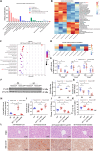
(A) Schematic representation of the generation of hepatic Nmnat1 knockout (Nmnat1-LKO) mice by crossing Nmnat1 Flox (Nmnat1-Ctrl) mice with Alb-Cre mice. (B) mRNA expressions of Nmnat1, Nmnat2, and Nmnat3 in the livers of 12-week-old male mice fed a normal chow diet (n = 8). (C to O) Nmnat1-Ctrl and Nmnat1-LKO mice were given a PF or AF diet for 8 weeks plus a single binge of ethanol (5 g/kg) before 4 hours of tissue collection. (C) Liver nuclear NAD+ content (n = 8). (D) Liver nuclear NADH content (n = 8). (E) Liver nuclear NAD+/NADH ratio (n = 8). (F) Plasma ALT levels (n = 8). (G) Plasma AST levels (n = 8). (H) Liver TG content (n = 8). (I) Liver H&E staining. (J) Liver Oil red O staining (n = 4). (K) mRNA levels of lipid-metabolizing enzymes in the mouse liver (n = 8). (L) Liver immunohistochemistry of MPO staining. (M) mRNA expressions of Il-1β and Cxcl1 in the mouse liver (n = 8). (N) Liver Sirius red staining. (O) mRNA expressions of Tgf-β and Acta2 in the mouse liver (n = 8). Data are presented as the means ± SD. *P < 0.05 represents statistical difference.
Fig. 4.
Fig. 4.. CSAD-regulated taurine metabolism is potentially involved in NMNAT1-regulated ALD.
(A and B) Nontargeted metabolomic KEGG analysis of the differential metabolite enrichment pathway (A) and differential metabolites in amino acid metabolic pathways (B) in AF Nmnat1-Ctrl and Nmnat1-LKO mouse livers (n = 4). (C and D) Transcriptome KEGG enrichment signal pathway analysis (C) and differential genes expression in taurine and hypotaurine metabolism signaling pathways (D) in AF Nmnat1-Ctrl and Nmnat1-LKO mouse livers (n = 4). (E) mRNA expressions of Csad, Fmo2, and Fmo3 in the mouse liver (n = 8). (F) CSAD protein expression in the mouse liver (n = 3). (G) Liver taurine content (n = 8). (H to L) Liver-specific CSAD overexpression mice were generated by caudal vein injection with an AAV8-constructed vector containing the Csad sequence. Mice injected with a null vector serve as the control. Taurine (1 g/kg per day) was supplemented in the diet of the AF + taurine group. (H) mRNA expressions of Csad in the mouse liver (n = 8). (I) Liver taurine content (n = 8). (J) Plasma ALT levels (n = 8). (K) Liver TG content (n = 8). (L) Liver H&E and Oil red O staining (n = 4). The protein band intensity was quantified by ImageJ. Data are presented as the means ± SD. *P < 0.05 represents statistical difference.
Fig. 5.
Fig. 5.. Nmnat1-LKO abrogates NMN supplementation–alleviated ALD.
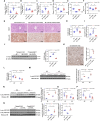
(A to J) Nmnat1-Ctrl and Nmnat1-LKO mice were supplemented with/without NMN (500 mg/kg per day) in the AF group. (A) Liver nuclear NAD+ content (n = 8). (B) Liver nuclear NADH content (n = 8). (C) Liver nuclear NAD+/NADH ratio (n = 8). (D) Plasma ALT levels (n = 8). (E) Plasma AST levels (n = 8). (F) Liver TG content (n = 8). (G) Liver H&E and Oil red O staining (n = 4). (H) Liver taurine content (n = 8). (I) mRNA expressions of Csad in the mouse liver (n = 8). (J) CSAD protein expression in the mouse liver (n = 4). (K to P) Liver immunohistochemistry of acetylated-lysine staining (K), SIRT1 activity (L), acetyl-FOXO1 protein expression (M), acetyl-HNF4α protein expression (N), and mRNA expressions of Cyp7a1 (O) and G6pc (P) was detected in AF Nmnat1-Ctrl and Nmnat1-LKO mouse livers (n = 3 to 8). (Q) Acetyl-FOXO1 and acetyl-HNF4α protein expressions were measured in the liver of NMN-administered Nmnat1-LKO ALD mice (n = 4). The protein band intensity was quantified by ImageJ. Data are presented as the means ± SD. *P < 0.05 represents statistical difference. MOD, mean optical density.
Fig. 6.
Fig. 6.. PPARα activation abrogates Nmnat1-LKO–aggravated ALD.
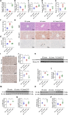
(A and B) Expression of PPARα protein (A) and mRNA expressions of Cpt1, Cpt2, and Acox1 (B) in AF Nmnat1-Ctrl and Nmnat1-LKO mouse livers (n = 3 to 8). (C to F) Nmnat1-Ctrl and Nmnat1-LKO mice were supplemented with/without Wy-14643 (10 mg/kg per day) in AF mice. (C) Plasma ALT levels (n = 8). (D) Plasma AST levels (n = 8). (E) Liver H&E and Oil red O staining (n = 4). (F) Liver TG content (n = 8). (G to K) Liver Pparα mRNA (G), PPARα protein (H), and Cpt1, Cpt2, and Acox1 mRNA [(I) to (K)] expressions were detected in the liver of AF liver–specific Csad-overexpressing or taurine-supplemented mice (n = 3 to 8). The protein band intensity was quantified by ImageJ. Data are presented as the means ± SD. *P < 0.05 represents statistical difference.
Fig. 7.
Fig. 7.. Liver-specific NMNAT1 overexpression ameliorates alcohol-induced hepatic steatosis.
Liver-specific Nmnat1 overexpression mice were generated by caudal vein injection with an AAV8-constructed vector containing the Nmnat1 sequence. Mice injected with a null vector serve as the control. (A) mRNA expressions of Nmnat1 in mice. (B) Liver nuclear NAD+ content (n = 8). (C) Liver nuclear NADH content (n = 8). (D) Liver nuclear NAD+/NADH ratio (n = 8). (E) Plasma ALT levels (n = 8). (F) Plasma AST levels (n = 8). (G) Liver TG content (n = 8). (H) Liver H&E, Oil red O, and MPO staining (n = 4). (I) Immunohistochemistry of acetylated-lysine staining (n = 4). (J) SIRT1 activity (n = 6). (K) Acetyl-HNF4α protein expression in the mouse liver (n = 4). (L) Liver taurine content (n = 8). (M) CSAD protein expression in the mouse liver (n = 4). (N) mRNA expressions of Csad in the mouse liver (n = 8). (O) PPARα protein expression in the mouse liver (n = 4). (P) mRNA expressions of Pparα in the mouse liver (n = 8). The protein band intensity was quantified by ImageJ. Data are presented as the means ± SD. *P < 0.05 represents statistical difference.
Fig. 8.
Fig. 8.. Schematic mechanistic illustration of hepatic NMNAT1–regulated ALD.

References

    1. Seitz H. K., Bataller R., Cortez-Pinto H., Gao B., Gual A., Lackner C., Mathurin P., Mueller S., Szabo G., Tsukamoto H., Alcoholic liver disease. Nat. Rev. Dis. Primers. 4, 16 (2018). - PubMed
    1. Mackowiak B., Fu Y., Maccioni L., Gao B., Alcohol-associated liver disease. J. Clin. Invest. 134, e176345 (2024). - PMC - PubMed
    1. Covarrubias A. J., Perrone R., Grozio A., Verdin E., NAD+ metabolism and its roles in cellular processes during ageing. Nat. Rev. Mol. Cell Biol. 22, 119–141 (2021). - PMC - PubMed
    1. French S. W., Chronic alcohol binging injures the liver and other organs by reducing NAD+ levels required for sirtuin’s deacetylase activity. Exp. Mol. Pathol. 100, 303–306 (2016). - PubMed
    1. Hao L., Sun Q., Zhong W., Zhang W., Sun X., Zhou Z., Mitochondria-targeted ubiquinone (MitoQ) enhances acetaldehyde clearance by reversing alcohol-induced posttranslational modification of aldehyde dehydrogenase 2: A molecular mechanism of protection against alcoholic liver disease. Redox Biol. 14, 626–636 (2018). - PMC - PubMed

Substances