. 2025 Sep:59:102449.
doi: 10.1016/j.tranon.2025.102449. Epub 2025 Jun 26.

PHF6 and RUNX1 mutations cooperate to accelerate leukemogenesis

Affiliations

PHF6 and RUNX1 mutations cooperate to accelerate leukemogenesis

Yueh-Chwen Hsu et al. Transl Oncol. 2025 Sep.

Abstract

Background: RUNX1 is a critical transcription factor in hematopoiesis and its mutations occur in various hematological diseases. PHF6 (plant homeodomain finger gene 6) is regarded as an epigenetic modifier, and its mutations are seen in myeloid and lymphoid leukemia. Previous studies have shown positive association of these two mutations. However, the joint pathological effects of these two genetic alterations remained unexplored.

Methods: We sought to investigate the pathological basis of the association between these two mutations. We first analyzed the clinical, genetic, and transcriptomic features of our cohort of patients with acute myeloid leuemia (AML) focusing on these two mutations. We transduced RUNX1 mutant into the genetically engineered Phf6 knockout (KO) mouse model to generate single- and double-mutated mice for in vivo experiments.

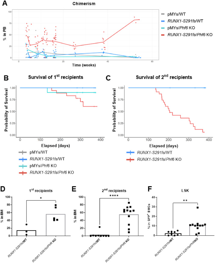
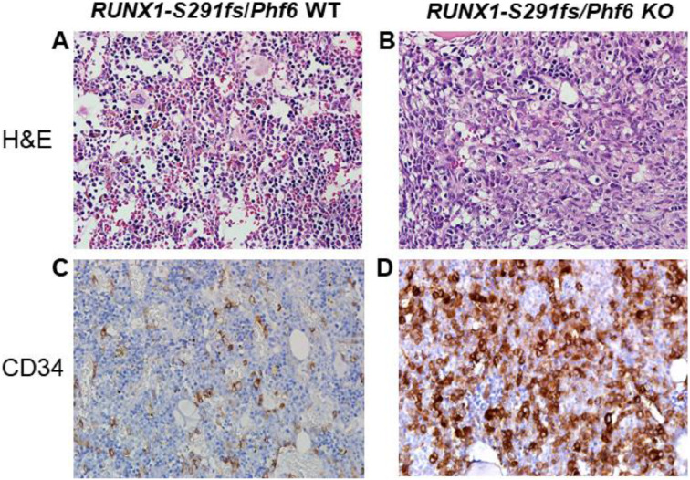
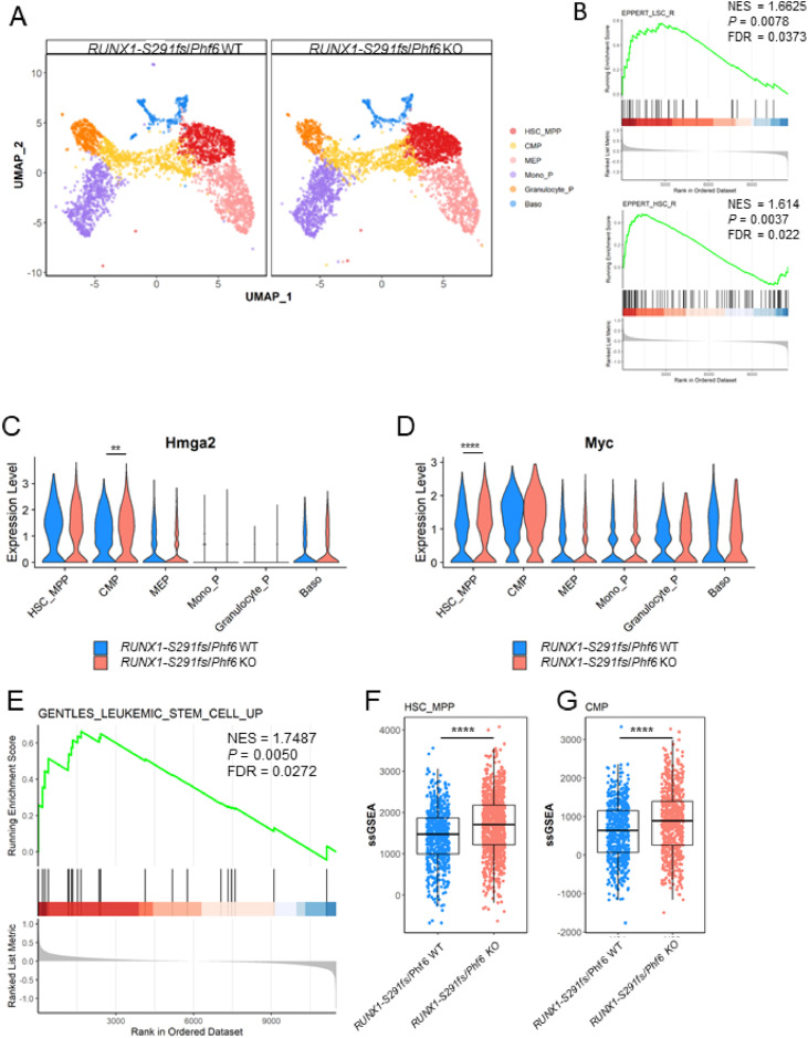
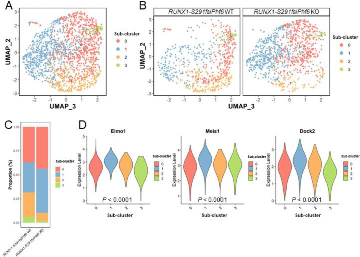
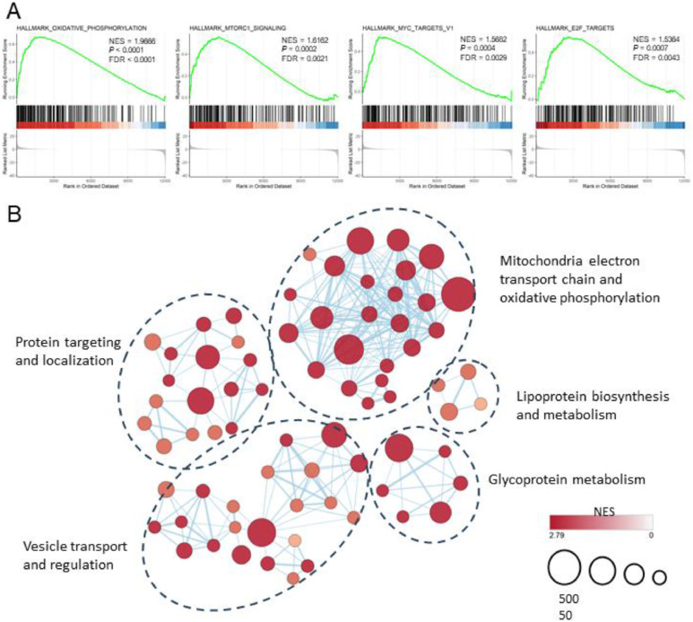
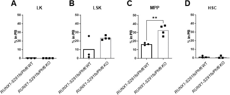
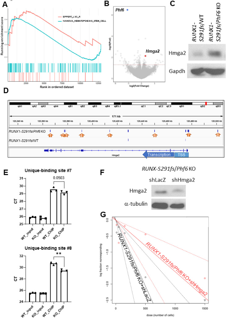
Results: In our 1188 adult AML patients, we observed frequent co-occurrence of PHF6 and RUNX1 mutations, and particularly worse clinical outcomes in these double-mutated patients. Double-mutated bone marrow (BM) cells displayed enriched leukemogenesis-related transcriptomic signatures and significantly higher engraftment capacity. The recipient mice transplanted with double-mutated BM cells developed AML with significantly shortened survival. Furthermore, we discovered that the multipotent progenitors (MPPs) were the main cell subpopulation responsible for double-mutated BM cell-induced leukemia. We noted significant up-regulation of high mobility group AT-hook 2 (Hmga2) in double-mutated MPPs and knock-down of Hmga2 abated the self-renewal capacity in vitro..

Conclusions: Our findings highlighted the synergistic leukemogenic potential of Phf6 and RUNX1 mutations in vivo, and provided insights into the molecular mechanisms accounting for this very high-risk disease entity.

Keywords: Acute myeloid leukemia; Hmga2; Leukemia stem cell; PHF6, RUNX1.


Conflict of interest statement

Declaration of competing interest The authors declare that they have no known competing financial interests or personal relationships that could have appeared to influence the work reported in this paper.

Figures

Fig 1
Fig. 1
Phf6 KO cells with RUNX1-S291fs overexpression exhibit a high reconstitution capacity. A Engraftment capacity was assayed in vivo by measuring the chimerism of various cell genotypes in mouse peripheral blood. The pMYs/WT (RUNX1 WT and Phf6 WT) and pMYs/Phf6 KO (RUNX1 WT and Phf6 KO) cells exhibited similar engraftment capacities. However, RUNX1-S291fs/Phf6 KO cells had higher engraftment capacity than RUNX1-S291fs/WT cells. Data are represented as the mean (pMYs/WT n = 5, RUNX1-S291fs/WT n = 16, pMYs/Phf6 KO n = 3, and RUNX1-S291fs/Phf6 KO n = 21; independent biological replicates). B Differences in overall survivial were modest between RUNX1-S291fs/Phf6 KO and RUNX1-S291fs/WT mice in first transplantation. C In the second transplantation, RUNX1-S291fs/Phf6-KO group had worse overall survival (P < 0.0001). Data are represented as the survival proportion (n = 11 and n = 23, respectively; independent biological replicates; statistical analysis via the Mental–Cox test). DRUNX1-S291fs/Phf6 KO cells had high chimerism in the BM in first transplantation at nine-month post-transplant (P = 0.0167). Data are represented as the mean (n = 3 and n = 5, respectively; independent biological replicates; *P < 0.05). ERUNX1-S291fs/Phf6 KO cells had high chimerism in the BM in second transplantation at six-month post-transplant (P < 0.0001). Data are represented as the mean (n = 9 and n = 12, respectively; independent biological replicates; statistical analysis via unpaired t-test; ****P < 0.0001). FRUNX1-S291fs/Phf6 KO cells had a high proportion of Lin-Sca-1+c-Kit+ (LSK) cells in the donor-derived BM in second transplantation (P = 0.0022). Data are representd as the mean (n = 9 and n = 12, respectively; independent biological replicates; statistical analysis via unpaired t-test; **P < 0.01).
Fig 2
Fig. 2
RUNX1-S291fs/Phf6 KO cells lead to severe blood diseases. A and B Hematoxylin and eosin (H&E) staining revealed increased blasts in the BM of RUNX1-S291fs/Phf6 KO mice. C and DRUNX1-S291fs/Phf6-KO mice had a significantly higher number of CD34+ blasts than the RUNX1-S291fs/WT mice.
Fig 3
Fig. 3
Single-cell transcriptomic profiling of RUNX1-S291fs/WT and RUNX1-S291fs/Phf6-KO murine hematopoietic stem and progenitor cells (HSPCs). A Uniform manifold approximation and projection (UMAP) representation of GFP+Lin- BM cells from RUNX1-S291fs/WT and RUNX1-S291fs/Phf6-KO mice. B Enrichment of LSC- (upper panel) and HSC-related (lower panel) signature of the HSC_MPP cluster in RUNX1-S291fs/Phf6 KO cells. Violin plots depicting the normalized expression levels of Hmga2C and MycD in each cluster. Data are represented as the violin plot (both n = 1; statistical analysis via Wilcoxon rank sum test; **P < 0.01; ****P < 0.0001). E Gene set enrichment analysis (GSEA) showed significant enrichment in the LSC geneset (GENTLES_LEUKEMIC_STEM_CELL_UP) in the RUNX1-S291fs/Phf6-KO versus RUNX1-S291fs/WT cells. Single-sample GSEA (ssGSEA) analysis revealed enrichment of GENTLES_LEUKEMIC_STEM_CELL_UP geneset in the HSC/MPP F and CMP G clusters of RUNX1-S291fs/Phf6-KO cells. Data are represented as the boxplot (both n = 1; statistical analysis via Wilcoxon rank sum test; ****P < 0.0001).
Fig 4
Fig. 4
Transcriptomic dissection of the HSC/MPP cluster. A UMAP representation of the sub-clusters within the parental HSC/MPP cluster. BRUNX1-S291fs/Phf6 KO cells exhibited a distinct sub-cluster composition compared to the RUNX1-S291fs/WT cells. C Proportions of sub-clusters 0 and 1 were higher in RUNX1-S291fs/Phf6 KO cells than in RUNX1-S291fs/WT cells. D Normalized expression levels of engulfment and cell motility 1 (Elmo1), myeloid ecotropic viral integration site 1 (Meis1), and dedicator of cytokinesis 2 (Dock2) were significantly higher in sub-cluster 1 than in other sub-clusters. Data are represented as the violin plot (both n = 1; statistical analysis via Kruskal–Wallis test).
Fig 5
Fig. 5
Gene expression profiling of RUNX1mut/PHF6mut acute myeloid leukemia (AML) patients. A Double-mutated patients exhibited enriched biological functions associated with oxidative phosphorylation, mTORC1, MYC, and E2F pathways. B Enrichment map of Gene Ontology terms demonstrating the significantly perturbed functions in double-mutated patients.
Fig 6
Fig. 6
Identification of the main cell population that contributed to RUNX1-S291fs/Phf6 KO cell-induced disease. A Transplantation of Lin-c-Kit+Sca-1- (LK) cells with both single- and double-mutated genotypes failed to restore hematopoiesis in the recepients. B Transplantation of LSK cells with both single- and double-mutated genotypes restored hematopoiesis in the recepients, but the chimerism was similar between the two groups. C Transplantation of RUNX1-S291fs/Phf6 KO MPPs resulted in a higher chimerism than that of RUNX1-S291fs/WT MPPs (P = 0.0062). Data are represented as the mean (n = 3 and n = 4, respectively; independent biological replicates; statistical analysis via unpaired t-test; **P < 0.001). D Both groups of HSCs showed low engraftment capacity.
Fig 7
Fig. 7
Gene expressiong profiling of RUNX1-S291fs/Phf6 KO MPPs. A GSEA showing the signatures of hematopoietic and leukemia stem cells enriched in RUNX1-S291fs/Phf6 KO MPPs. B Volcano plot of differentially expressed genes between RUNX1-S291fs/Phf6 KO and RUNX1-S291fs/WT MPPs; red dotted line indicates P = 0.05. Expression of Hmga2 was high in RUNX1-S291fs/Phf6nKO MPPs (P = 0.0075). Data are represented as the volcano plot (both n = 4; independent biological replicates; statistical analysis performed with the limma method). C Western blotting indicated that RUNX1-S291fs/Phf6 KO mouse BM cells had higher Hmga2 level compared to RUNX1-S219fs/WT ones. D RUNX1-S291fs had eight unique-binding sites, denoted by arrows, at the Hmga2 locus in RUNX1-S291fs/Phf6 KO cells, and two of these binding sites were located upstream the TSS region. In contrast, no unique binding sites were identified for RUNX1-S291fs in RUNX1-S291fs/WT cells. E Chromatin immunoprecipitation (ChIP)-quantitative polymerase chain reaction (qPCR) verification of the unique-binding peaks upstream the TSS region in the Hmga2 locus. Data are represented as the mean (both n = 3; technical replicates; statistical analysis via a paired t-test; **P < 0.01). The lower the CT levels, the higher the amount of templates was. F Western blotting results corroborating Hmga2 downregulation in RUNX1-S291fs/Phf6 KO mouse BM cells transduced with either control shRNA (shLacZ) or Hmga2 shRNA (shHmga2). G LTC-IC assay revealed that RUNX1-S291fs/Phf6 KO cells with shLacZ (black line) had a much higher long-term colony-forming capacity than the RUNX1-S291fs/Phf6 KO cells with shHmga2 (red line; P = 0.0406). Data presented as a trend line represent the estimated active cell frequency, and dotted lines indicate the 95 % confidence interval in each group (RUNX1-S291fs/Phf6 KO+shLacZ n = 30 and RUNX1-S291fs/Phf6 KO+shHmga2 n = 30; independent technical replicates; statistical analysis via Chi-square test). The down-pointing triangle at cell dose of 1500 cells means that all wells at this dose level formed colonies.

Similar articles

References

    1. Hsu Y.-C., Chen T.-C., Lin C.-C., Yuan C.-T., Hsu C.-L., Hou H.-A., et al. Phf6-null hematopoietic stem cells have enhanced self-renewal capacity and oncogenic potentials. Blood Adv. 2019;3(15):2355–2367. - PMC - PubMed
    1. Miyagi S., Sroczynska P., Kato Y., Nakajima-Takagi Y., Oshima M., Rizq O., et al. The chromatin-binding protein Phf6 restricts the self-renewal of hematopoietic stem cells. Blood. 2019;133(23):2495–2506. Jun 6. - PubMed
    1. Wendorff A.A., Quinn S.A., Rashkovan M., Madubata C.J., Ambesi-Impiombato A., Litzow M.R., et al. Phf6 loss enhances HSC self-renewal driving tumor initiation and leukemia stem cell activity in T-ALL. Cancer Discov. 2019;9(3):436–451. - PMC - PubMed
    1. McRae H.M., Garnham A.L., Hu Y., Witkowski M.T., Corbett M.A., Dixon M.P., et al. PHF6 regulates hematopoietic stem and progenitor cells and its loss synergizes with expression of TLX3 to cause leukemia. Blood. 2019;133(16):1729–1741. Apr 18. - PMC - PubMed
    1. Yuan S., Wang X., Hou S., Guo T., Lan Y., Yang S., et al. PHF6 and JAK3 mutations cooperate to drive T-cell acute lymphoblastic leukemia progression. Leukemia. 2022;36(2):370–382. Feb. - PMC - PubMed