Review
. 2025 Jul 1;37(7):koaf150.
doi: 10.1093/plcell/koaf150.

How meristems shape plant architecture in cereals-Cereal Stem Cell Systems (CSCS) Consortium

Affiliations
Review

How meristems shape plant architecture in cereals-Cereal Stem Cell Systems (CSCS) Consortium

Thomas Dresselhaus et al. Plant Cell. .

Abstract

Meristems are major determinants of plant architecture, diversification, and acclimation to environmental stresses. Moreover, meristems play also a major role during crop domestication and are fundamentally important for the productivity of crop plants as they directly determine biomass and grain yield. While vegetative meristems shape the basic plant body plan and produce all above- and below-ground parts of plants, some vegetative meristems transit to reproductive meristems, forming sexual organs and germ cells. Most knowledge about plant meristems was generated using the model plant Arabidopsis. Compared with Arabidopsis, architecture of grass or cereals, including crops like maize, wheat, barley, rice and sorghum, is more complex: cereals produce additional organs like a coleoptile, seminal roots originating from the scutellar nodes in the embryo and shoot-borne crown roots as well as highly complex inflorescence meristems with meristem types absent in eudicots. Moreover, studies in cereals indicated that paradigms based on studies using Arabidopsis are not universally applicable. This review therefore aims to provide a comprehensive overview about the initiation, establishment, maintenance, and function of the various cereal meristems and their stem cell niches that shape our most important crop plants. Stem cell-like systems involved in leaf pattering and germline formation are also considered. The focus is also on the significant progress that has been made recently using novel tools to elucidate gene regulatory networks underlying the development and function of the various cereal meristems.


Conflict of interest statement

Conflict of interest statement. None declared.

Figures

Figure 1.
Figure 1.
Meristem initiation and organ formation during cereal embryogenesis. Note that stages are not drawn to scale. A) Embryonic structures of Arabidopsis compared with different cereals. Shoot and root apical meristems are indicated. Partly front and side views are shown. B) Gene expression pattern regulating cell fate determination during early embryogenesis in cereals with maize as an example. Embryonic stages are indicated. Different colors mark embryonic cells expressing indicated genes. C) Auxin and CK responses during maize embryogenesis. Auxin flux is represented by black arrows. The DR5 and TCS reporter activities are marked in saxe-blue and pink, respectively. Abbreviations: Col, coleoptilar; Ep, epiblast; Hy, hypocotyl; Lp, leaf primordium; Ram, root apical meristem; Sam, shoot apical meristem; Sc, scutellum; Su, suspensor; Va, vasculature. C) Adapted from Chen et al. (2014).
Figure 2.
Figure 2.
Organization of the vegetative SAM and the pathways controlling their activity in maize as a cereal model. A) The SAM is organized into discrete, functional domains, shown on the schematic on the left. Color-coded is a list of genes (see text for details) whose expression corresponds to functionally distinct domains in the SAM. Abbreviations: CZ, central zone; L1, epidermal layer; L2, inner cell layers; OC, putative organizing center; P0, incipient primordium; P1, primordium; PZ, peripheral zone. B) Simplified view of CLE-peptide mediated regulation of meristem activity. Multiple CLE-peptides are perceived by Leucine-Rich Repeat CLAVATA receptors and often negatively regulate WOX gene expression. C) Overview of the hormonal regulation of SAM homeostasis (see text for gene names). The schematic depicts known interactions between CK and auxin signaling pathways during SAM regulation. Red arrows represent metabolic pathways, while black arrows represent genetic interactions (full, validated interaction; dotted, putative interaction).
Figure 3.
Figure 3.
Root apical meristems (RAMs) in crops. A to D) Schematic overview of different cell types in the RAMs of Arabidopsis (A), rice (B), barley (C) and maize (D) as indicated. Abbreviations: CEI, cortex/endodermis initials; CSC, columella stem cells; LRC/EI, lateral root cap/epidermis initials; QC, quiescent center; SI, stele initials. A′ to D′) Expression patterns of PLETHORA 1 (PLT1), SCARECROW (SCR), SHORTROOT (SHR), WUSCHEL-RELATED HOMEOBOX 5/QHB (WOX5/QHB) and their overlapping pattern as indicated in the RAMs of Arabidopsis (A′), rice (B′), barley (C′) and maize (D′). Auxin flow is indicated with gray arrows.
Figure 4.
Figure 4.
Control of phytomer initiation and ICM activation. The main shoot of vascular plants (barley as a showcase) consists of several repeated phytomers, which originate from the SAM. A gradual shift in the outgrowth of 2 lateral organs—the leaf and the axillary meristem (AxM)—marks the transition from vegetative (Veg) to reproductive (Rep) growth. In cereals, the AxM becomes a spikelet meristem (SM) in reproductive growth. The antagonistic YABBY–KNOX1 regulatory module determines nodes and internodes in a phytomer, which also includes a foot (non-elongating domain at the base) and the ICM. Cell division of the ICMs then displaces cells upward to form an internode (IntN). TFs are highlighted that can respond to various environmental stressors to activate the ICM and induce internode elongation. la, lacuna; —> activation; ——| repression; ------| negative correlation.
Figure 5.
Figure 5.
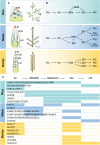
Differences in IMs of cereals. A) Schematic illustrations of immature and mature inflorescences of rice, maize, and barley, respectively. The immature inflorescence of rice includes the IM and BM), while the mature inflorescence displays the primary branches (PB), secondary branches (SB), and spikelets. In maize, the immature ear inflorescence shows the IM, spikelet meristem (SM), and SPM, whereas the mature tassel inflorescence depicts the long branches (indicated by side arrows) with the paired spikelets. The immature inflorescence of barley demonstrates the progression of reproductive meristems, encompassing the IM, TSM, 3 SMs, and 3 FMs from the apex to the base. In contrast, the mature inflorescence exhibits 2 rows of spikelets. B) Developmental trajectories from vegetative meristem (VM) to FM in rice, maize, and barley are illustrated. C) A common differentiation pathway from cereal IM to FM is shown, followed by the genes expressed in various meristems along the pathway for rice (cyan blue), maize (blue), and barley (orange).
Figure 6.
Figure 6.
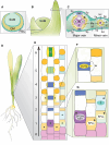
Stem cell systems during leaf development. A) Schematic of a cross-section through a young leaf primordium with the inner ground meristem (GM) and outer protodermal stem cells (PSC). The primordium envelops the vegetative SAM. B) Schematic of the SAM with a young (left) and an older (right) leaf primordium. C) Schematic of a cross-section through the mature leaf zone with a major and minor vein. Vein tissues include xylem (X), phloem (P), mestome sheath (M) and bundle sheath cells (B). Sclerenchymatic cells (Scl) are indicated above and below the veins and the outermost cell layers are epidermal cells with stomatal pores (Sp) adjacent to the veins. D) Schematic of a young grass seedling. E) Schematic of the leaf epidermal developmental zone. Seven developmental stages are shown with protodermal cells (PSCs, stage 0), and the 6 stages of stomatal development from lineage establishment to mature stomatal complexes (stage 1–6); asterisks indicate cells that retain division capacity beyond the PSC stage (0). F) Magnification of the stomatal subsidiary mother cell (SMC, asterisk). The TF BdMUTE is expressed in the guard mother cell (GMC, blue) and moves into lateral neighbor cells to establish SMC identity and induce an asymmetric division to form subsidiary cells (SCs, indicated by dashed line). G) Magnification of the developmental stages 0 to 2. Cells with an asterisk divide asymmetrically in transverse orientation to form stomatal precursors, hair cell precursors or silica cell precursors, respectively. The different TFs specifying cell file identities are indicated.
Figure 7.
Figure 7.
Female germline specification. Sketch of megaspore mother cell (MMC) formation in maize (A to C) and key pathways involved in MMC specification in Arabidopsis (D and E). A) The archesporium is formed at the nucellar region of the ovule primordium and likely involves a stem cell-like system. B) Only 1 subepidermal cell per ovule primordium acquires the reproductive fate, the archesporial cell (AR). Adjacent to the AR, the ovule primordium contains several cell types which presumably support the developing germline. C) The archesporial cell develops into a female meiocyte, i.e. the MMC. Surrounding cell types are indicated. D) Ovule primordium of Arabidopsis containing an MMC. Additional cell types can be distinguished. E) Key pathways involved in MMC specification in Arabidopsis: the upper epidermal layer promotes the establishment of an environment suitable for MMC formation, while the lower epidermal layer together with the companion cells suppresses MMC identity. Pathways to acquire and maintain MMC identity are also indicated. See text for details of molecular players and pathways.
Figure 8.
Figure 8.
Developmental progression and regulatory networks in male germline specification. A) Schematic representation of anther development in maize as an example. Early stages involve the differentiation of L1 and L2-d layers (anther length 90 µm), with the emergence of archespore cells (ARs) at an anther length of 120 µm. Subsequent stages lead to the formation of somatic layers, including the tapetum (TAP), middle layer (ML), endothecium (EN), epidermis (EP), and vasculature (VA), shown in cross-section at 1,200 µm length stage. B) Comparative lineage relationships of maize and Arabidopsis anther tissues. L1-d and L2-d layers give rise to different cell types, including the epidermis, primary and secondary parietal cell layers (PPL, SPL), and AR, which later become pollen mother cells (PMC). Note that the formation of somatic anther lobe layers (TAP, ML, EN) has been described by different models in both species. The additional L3-d cells in Arabidopsis contribute to VA and connective (CO) tissues. C) Regulators of anther development in maize and rice. See text for details.

Similar articles

References

    1. Abe M, Kobayashi Y, Yamamoto S, Daimon Y, Yamaguchi A, Ikeda Y, Ichinoki H, Notaguchi M, Goto K, Araki T. FD, a bZIP protein mediating signals from the floral pathway integrator FT at the shoot apex. Science. 2005:309(5737):1052–1056. 10.1126/science.1115983 - DOI - PubMed
    1. Abrash E, Anleu Gil MX, Matos JL, Bergmann DC. Conservation and divergence of YODA MAPKKK function in regulation of grass epidermal patterning. Development. 2018:145(14):dev165860. 10.1242/dev.165860 - DOI - PMC - PubMed
    1. Adamowski M, Friml J. PIN-dependent auxin transport: action, regulation, and evolution. Plant Cell. 2015:27(1):20–32. 10.1105/tpc.114.134874 - DOI - PMC - PubMed
    1. Ahmar S, Hensel G, Gruszka D. CRISPR/Cas9-mediated genome editing techniques and new breeding strategies in cereals—current status, improvements, and perspectives. Biotechnol Adv. 2023:69:108248. 10.1016/j.biotechadv.2023.108248 - DOI - PubMed
    1. Ahn JH, Miller D, Winter VJ, Banfield MJ, Lee JH, Yoo SY, Henz SR, Brady RL, Weigel D. A divergent external loop confers antagonistic activity on floral regulators FT and TFL1. EMBO J. 2006:25(3):605–514. 10.1038/sj.emboj.7600950 - DOI - PMC - PubMed

MeSH terms