

Mitral Annular Disruptions
- PMID: 40579097
- PMCID: PMC12273815
- DOI: 10.1016/j.jaccas.2025.104342
Mitral Annular Disruptions
Abstract
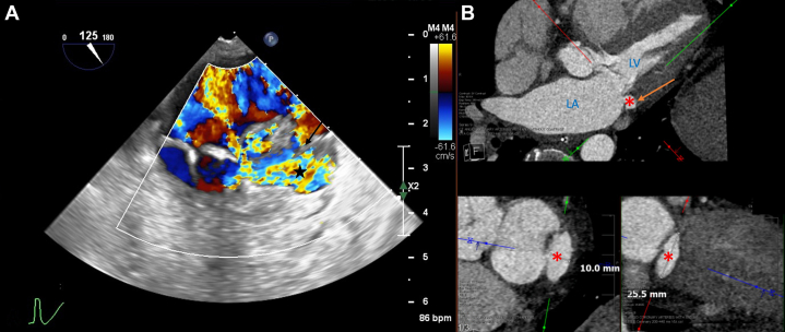
Background: Mitral annular disruption is increasingly recognized in our clinical practice and is most commonly due to infective endocarditis or transcatheter or hybrid balloon-expandable mitral valve deployment in the setting of mitral annular calcification.
Case summary: A case series is presented of 4 patients with mitral annular disruption in 4 distinct clinical scenarios, each requiring multimodality imaging for diagnosis.
Discussion: These defects are often difficult to detect on transthoracic imaging and challenging to interpret on transesophageal echocardiography given the unique flow pattern and a general unfamiliarity among echocardiographers with such a defect.
Take-home messages: These 4 cases describe mitral annular disruption caused by infective endocarditis or mitral valve replacement in the setting of mitral annular calcification. These cases stress the importance of careful multimodality cardiac imaging and maintaining a high clinical suspicion for mitral annular disruption, as it is often challenging to diagnose.
Keywords: endocarditis; mitral valve replacement.
Copyright © 2025 The Authors. Published by Elsevier Inc. All rights reserved.
Conflict of interest statement
Funding Support and Author Disclosures Dr Inglessis-Azueja has been a consultant for Edwards Lifesciences. Dr Foldyna has reported institutional research support from the National Institutes of Health/National Heart, Lung, and Blood Institute, AstraZeneca, MedImmune, Cleerly, and MedTrace. Dr Langer has been a consultant for Edwards Lifesciences. Dr Paras has received travel support from the Infectious Diseases Society of America and grant support from the Centers for Disease Control and Prevention. All other authors have reported that they have no relationships relevant to the contents of this paper to disclose.
Figures


References
-
- Guler A., Karabay C.Y., Gursoy O.M., et al. Clinical and echocardiographic evaluation of mitral valve aneurysms: a retrospective, single center study. Int J Cardiovasc Imaging. 2014;30(3):535–541. - PubMed
-
- Du Toit H.J., Von Oppell U.O., Hewitson J., Lawrenson J., Davies J. Left ventricular sub-valvar mitral aneurysms. Interact Cardiovasc Thorac Surg. 2003;2(4):547–551. - PubMed
-
- Pasic M., Unbehaun A., Buz S., Drews T., Hetzer R. Annular rupture during transcatheter aortic valve replacement: classification, pathophysiology, diagnostics, treatment approaches, and prevention. JACC Cardiovasc Interv. 2015;8(1 Pt A):1–9. - PubMed
Publication types
LinkOut - more resources
Full Text Sources
