Case Reports
. 2025 Jun 25;30(16):103761.
doi: 10.1016/j.jaccas.2025.103761.

Embolic Stroke Due to a Large Noncoronary Sinus of Valsalva Aneurysm: A Multimodality Imaging Diagnosis

Affiliations
Case Reports

Embolic Stroke Due to a Large Noncoronary Sinus of Valsalva Aneurysm: A Multimodality Imaging Diagnosis

Tyler C Miller et al. JACC Case Rep. .

Abstract

Background: A sinus of Valsalva aneurysm (SoVA) is a rare cardiac condition caused by the dilation of a coronary sinus. If untreated, it can commonly lead to valvular dysfunction, arrhythmias, or rupture.

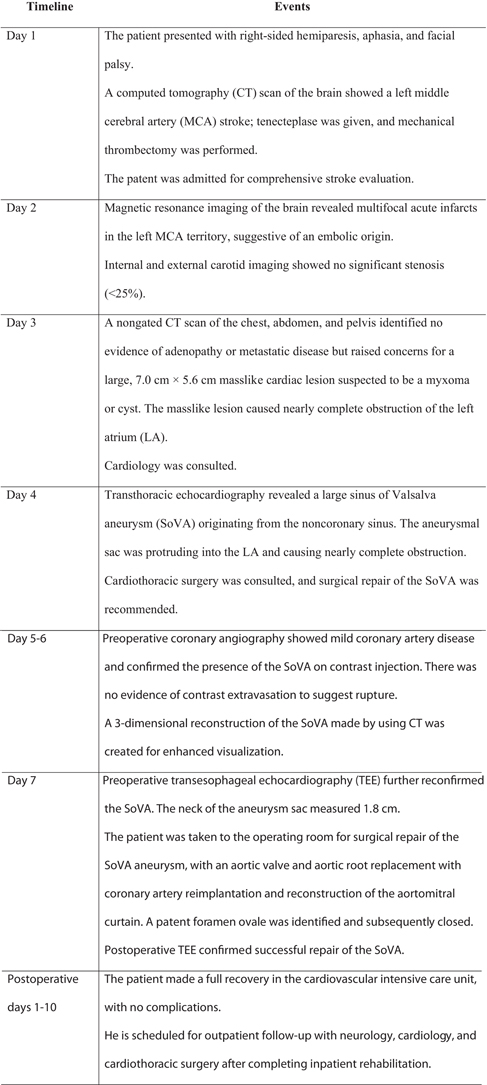
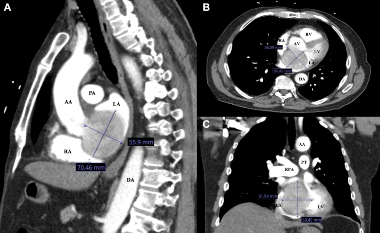
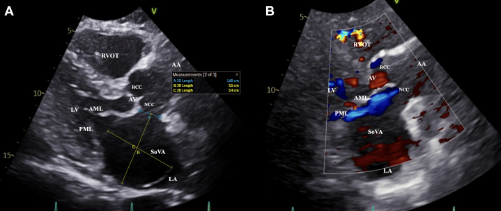
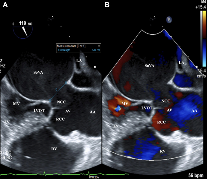
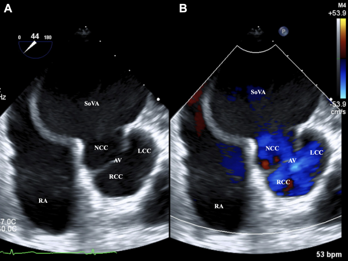
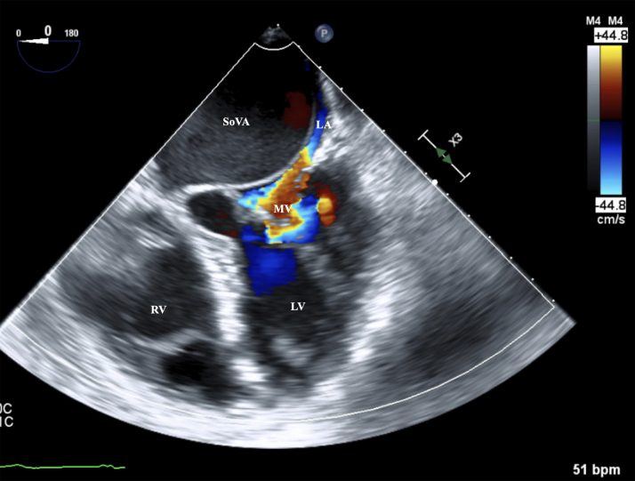
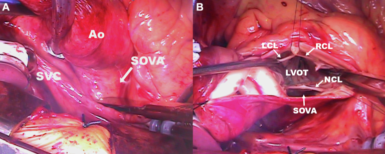
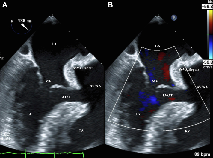
Case summary: A 71-year-old patient with hypertension and hyperlipidemia presented with an embolic stroke. Multimodality imaging revealed a large, 7.0 cm × 5.6 cm SoVA originating from the noncoronary sinus and causing nearly complete obstruction of the left atrium. The aneurysm was surgically repaired, and the patient made a full recovery.

Discussion: In rare cases, a stroke may be the initial presentation of a SoVA. The probable cause of the patient's stroke was attributed to thrombus formation within the SoVA that embolized.

Take-home messages: This case emphasizes the importance of multimodality imaging for the diagnosis of a SoVA and for planning surgical repair. Additionally, clinicians should consider a SoVA in the differential diagnosis for a patient presenting with a stroke.

Keywords: aortic root repair; computed tomography; coronary angiography; multimodality cardiac imaging; noncoronary sinus; sinus of Valsalva aneurysm; stroke; transesophageal echocardiogram; transthoracic echocardiogram.


Conflict of interest statement

Funding Support and Author Disclosures The authors have reported that they have no relationships relevant to the contents of this paper to disclose.

Figures

None
Graphical abstract
Figure 1
Figure 1
Initial Nongated Computed Tomography of the Chest in Sagittal, Axial, and Coronal Views Findings concerning for a large 7.0 cm × 5.6 cm masslike cardiac lesion suspected to be a myxoma or a cyst in (A) sagittal, (B) axial, and (C) coronal views. The masslike lesion caused nearly complete obstruction of the left atrium (LA). AA = ascending aorta; AV = aortic valve; DA = descending aorta; LV = left ventricle; PA = pulmonary artery; PT = pulmonary trunk; RA = right atrium; RPA = right pulmonary artery; RV = right ventricle.
Figure 2
Figure 2
Initial Transthoracic Echocardiography in the Parasternal Long-Axis View With Color Doppler A large sinus of Valsalva aneurysm (SoVA) originating from the noncoronary sinus, protruding into the left atrium (LA), and causing nearly complete obstruction. (A) The aneurysm measured 5.5 cm × 5.4 cm, and the aneurysm sac measured 1.6 cm on the parasternal long-axis view. (B) A communication is shown from the noncoronary sinus into the aneurysmal sac by using color Doppler. AML = anterior mitral leaflet; AV = aortic valve; NCC = noncoronary cusp; PML = posterior mitral leaflet; RCC = right coronary cusp; RVOT = right ventricle outflow tract; other abbreviations as in Figure 1.
Figure 3
Figure 3
Preoperative Transesophageal Echocardiography in the Midesophageal Long-Axis View With Color Doppler A large sinus of Valsalva aneurysm (SoVA) originating from the noncoronary sinus, protruding into the left atrium (LA), and causing nearly complete obstruction. (A) The large aneurysm sac measured 1.8 cm. (B) A communication is shown from the noncoronary sinus into the aneurysmal sac by using color Doppler. bpm = beats/min; MV = mitral valve; LVOT = left ventricular outflow tract; other abbreviations as in Figures 1 and 2.
Figure 4
Figure 4
Preoperative Transesophageal Echocardiography in the Midesophageal Aortic Valve Short-Axis View With Color Doppler (A) A large sinus of Valsalva aneurysm (SoVA) originating from the noncoronary sinus. (B) A communication is shown from the noncoronary sinus into the aneurysmal sac by using color Doppler. LCC = left coronary cusp; other abbreviations as in Figures 1 and 2.
Figure 5
Figure 5
Preoperative Transesophageal Echocardiography in the Midesophageal 4-Chamber View With Color Doppler The figure shows a large sinus of Valsalva aneurysm originating from the non-coronary sinus protruding into the left atrium, causing a near-complete obstruction on preoperative transesophageal echocardiogram in mid-esophageal four-chamber view. LA = left atrium; SoVA = sinus of Valsalva aneurysm. Abbreviations as in Figures 1 and 3.
Figure 6
Figure 6
Intraoperative Images of the SoVA (A) Findings of a large sinus of Valsalva aneurysm (SoVA). (B) A transverse aortotomy was performed and the aortic valve was exposed, revealing a 7-cm aneurysm behind the aorta (Ao) and severely compressing the left atrium. LCL = left coronary leaflet; LVOT = left ventricular outflow tract; NCL = noncoronary leaflet; RCL = right coronary leaflet; SVC = superior vena cava.
Figure 7
Figure 7
Postoperative Transesophageal Echocardiography in the Midesophageal Long-Axis View With Color Doppler (A) The surgically repaired sinus of Valsalva aneurysm (SoVA). (B) A communication is no longer present after surgical repair using color Doppler. Abbreviations as in Figure 1, Figure 2, Figure 3.

Similar articles

References

    1. Arcario M.J., Lou S., Taylor P., et al. Sinus of Valsalva aneurysms: a review with perioperative considerations. J Cardiothorac Vasc Anesth. 2021;35(11):3340–3349. - PMC - PubMed
    1. Takach T.J., Reul G.J., Duncan J.M., et al. Sinus of Valsalva aneurysm or fistula: management and outcome. Ann Thorac Surg. 1999;68(5):1573–1577. - PubMed
    1. Dev V., Goswami K.C., Shrivastava S., et al. Echocardiographic diagnosis of aneurysm of the sinus of Valsalva. Am Heart J. 1993;126:930–936. - PubMed
    1. Feldman D.N., Roman M.J. Aneurysms of the sinuses of Valsalva. Cardiology. 2006;106:73–81. - PubMed
    1. Fishbein M.C., Obma R., Roberts W.C. Unruptured sinus of Valsalva aneurysm. Am J Cardiol. 1975;35:918–922. - PubMed

Publication types

LinkOut - more resources